
|
Fig. 4
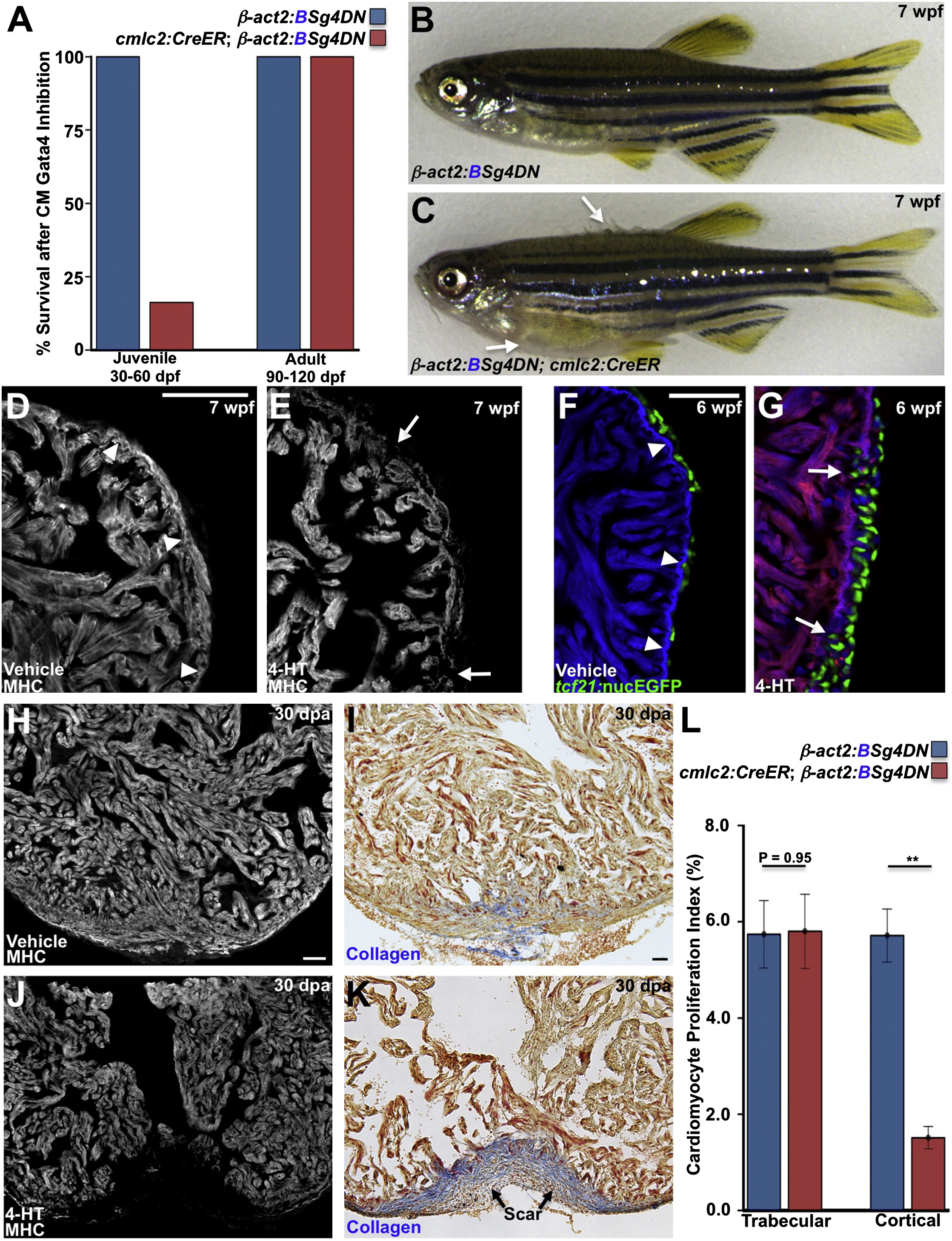
Gata4 Inhibition Blocks Juvenile Cortical Layer Formation and Adult Heart Regeneration (A) Survival of zebrafish after induction of a dominant-negative Gata4 construct (g4DN) for 30 days in cardiomyocytes at either 30 or 90 dpf. g4DN induction at 30 dpf sharply reduced survival of juveniles (n = 23–43), whereas g4DN induction at 90 dpf did not affect survival (n = 8–16). (B) A Cre(–) control animal exposed to 4-HT at 5 wpf, appearing normal at 7 wpf. (C) A Cre(+) clutchmate exposed to 4-HT at 5 wpf, with flared scales indicative of edema (arrows). (D and E) Sections of the 7 wpf ventricular wall after g4DN induction at 5 wpf, stained for myosin heavy chain (MHC). Control animals have cortical muscle at the base of the ventricle (arrowheads), whereas the wall is thinned and disrupted after g4DN induction (n = 4) (arrows). (F and G) Maturing juvenile ventricles visualized for tcf21:nucEGFP+ epicardium (green), g4DN (red), and β-actin2:BFP (blue). The 6 wpf control ventricle (F) has a contiguous wall (arrowheads) and normal epicardial cell distribution. g4DN induction at 5 wpf results in wall gaps (arrows) and increased epicardial cell presence at 6 wpf (G) (n = 6–9). (H–K) g4DN was induced in adults, and ventricular apices were resected, before analysis of regeneration at 30 dpa. Control animals (H and I) regenerated muscle with little or no scarring (blue indicates collagen), whereas g4DN induction blocked muscle regeneration (J) and caused scarring (K). (L) Quantification of proliferation of trabecular and cortical cardiomyocytes in 7 dpa ventricles of g4DN-expressing animals versus controls (n = 12–15). **p < 0.005, Student’s t test. Scale bars represent 50 μm. See also Figure S4 and Movies S2 and S3.