
|
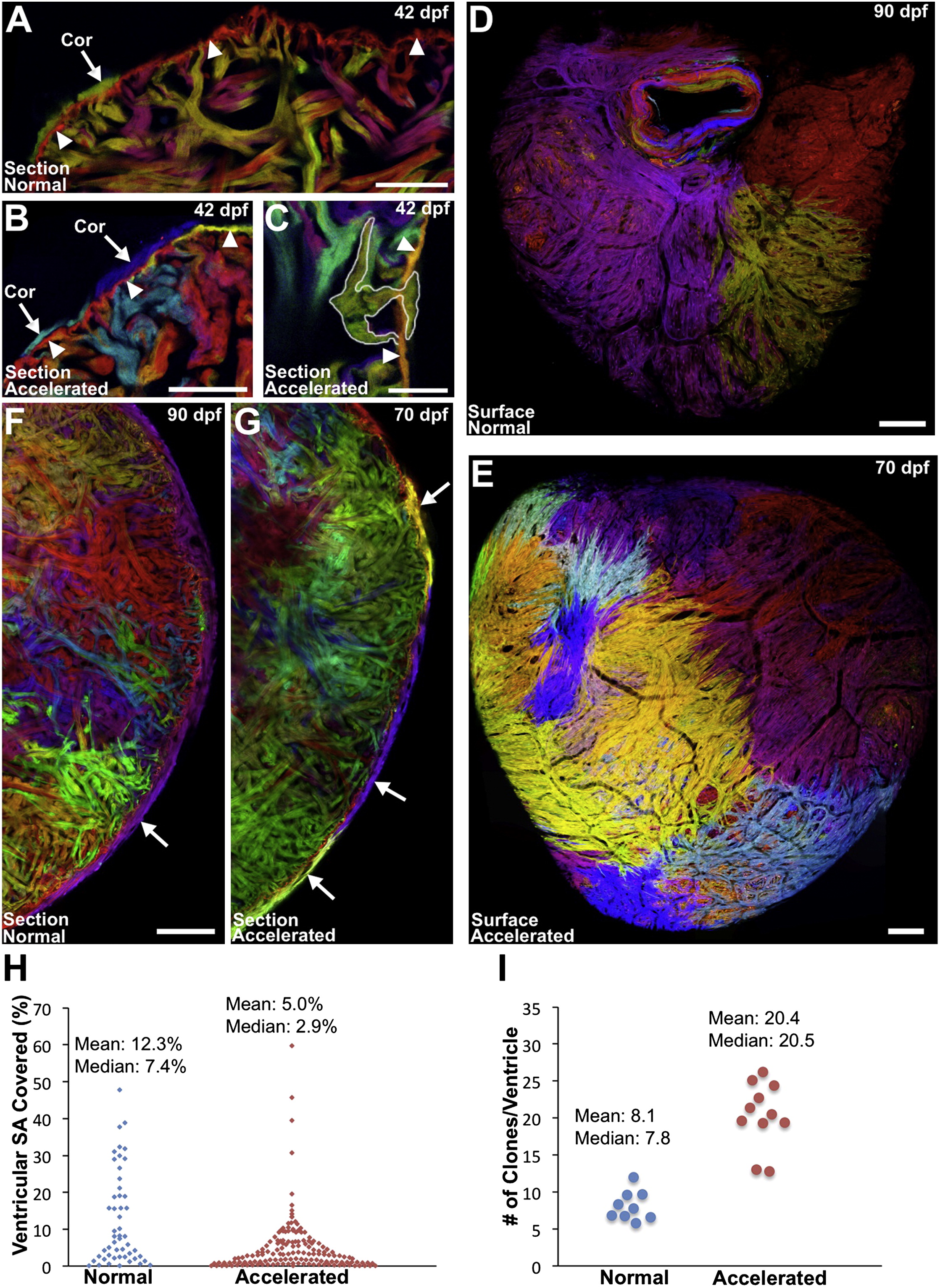
Fig. 3
Accelerating Growth Leads to Increased Contributions to the Cortical Layer (A–C) Sections of 6 wpf ventricles of cmlc2:CreER; priZm zebrafish that were labeled at 2 dpf and grown at normal (A) or accelerated growth (B and C) conditions. NG fish contained one or two small cortical clones at the base of the ventricle (A, arrow) outside the primordial layer (arrowheads). Multiple small cortical clones were seen in AG fish (B, arrows), including those breaching (C, outlined) the primordial layer (B and C, arrowheads). (D and E) Surface muscle of adult cmlc2:CreER; priZm ventricles after normal (D) or accelerated growth (E) conditions. More cortical clones are generated during AG. (F and G) Confocal slices through ventricles after normal (F) or accelerated (G) growth, indicating a higher number of cortical clones (arrows) after accelerated growth. (H) Percentage surface area (SA) occupied by clones after normal (51 clones, 9 ventricles) or accelerated (153 clones, 11 ventricles) growth. (I) Extrapolated number of surface clones per ventricle from data in (H). Scale bars represent 100 μm (C–G), 50 μm (A and B), and 25 μm (C). See also Figure S3.