
|
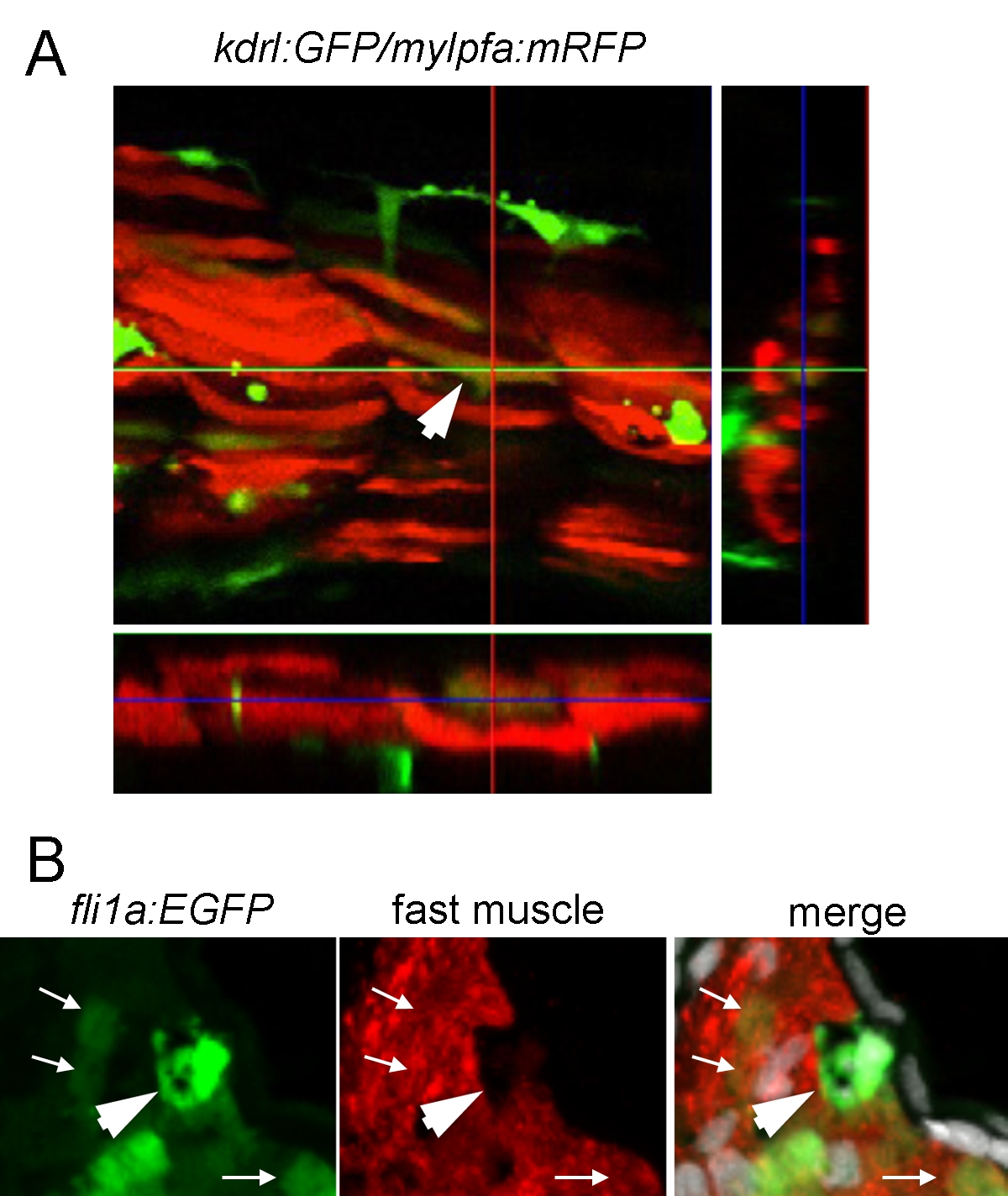
Fig. S8
Fast muscle–specific mylpfa:mRFP is co-expressed with kdrl:GFP following overexpression of Etv2. (A) Confocal image of the trunk of a kdrl:GFP/mylpfa:mRFP/hsp70l:etv2 triple transgenic embryo heat shocked at 24 hpf and imaged at 12 h post–heat shock. A GFP/mRFP double positive muscle fiber is highlighted by the arrow and x-axis and y-axis z-plane projections are presented below and to the right of the image respectively. (B) Fast muscle myosin and GFP colocalize in the trunk of kdrl:GFP/hsp70l:etv2 transgenic fish heat shocked at 24 hpf and imaged at 48 hpf. Red muscle fibers colocalize with GFP (white arrows). A strongly GFP positive cell located where a muscle fiber normally would be is negative for fast muscle myosin (large white arrowhead), suggesting this cell has lost its muscle cell identity.