
|
Fig. S1
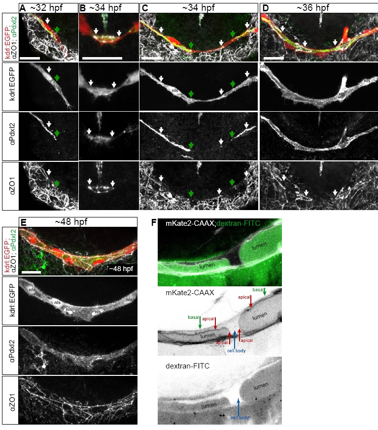
Podocalyxin-like 2 (Pdxl2) staining labels apical/luminal membrane during PLA fusion. Related to Fig. 1 and 2.
A-E. Antibody staining for Pdxl2 (apical, green) and ZO-1 (junctional, white), in the PLA sprouts of Tg(kdrl:EGFP)s843 embryos (red). Pdxl2 labels the invaginating apical membrane (A, green arrows) extending beyond junctional contacts in the PLA sprout (A, white arrow). During fusion, apical membrane is present within the new junctional ring (B, arrows). Apical membrane (C, green arrows) extends inside the tip cells after new contact formation (C, middle white arrow). After transcellular lumen formation, continuous apical surface in the PLA marks perfused lumen; the staining is present within unicellular tubes, connected by junctional rings (D white arrows). Continuous apical surface is present during subsequent cell rearrangements leading to formation of a multicellular tube (E). Related to Fig. 1.
F. Close view of the apical membrane invagination in a transgenic embryo TgBAC(kdrl:mKate2-CAAX)ubs16 (cell membranes in white) with dextran-FITC angiography (green). Basal and apical membrane compartments, cell body and lumen are marked in the single channel image. Related to Fig. 2.
Reprinted from Developmental Cell, 25(5), Lenard, A., Ellertsdottir, E., Herwig, L., Krudewig, A., Sauteur, L., Belting, H.G., and Affolter, M., In Vivo analysis reveals a highly stereotypic morphogenetic pathway of vascular anastomosis, 492-506, Copyright (2013) with permission from Elsevier. Full text @ Dev. Cell