
|
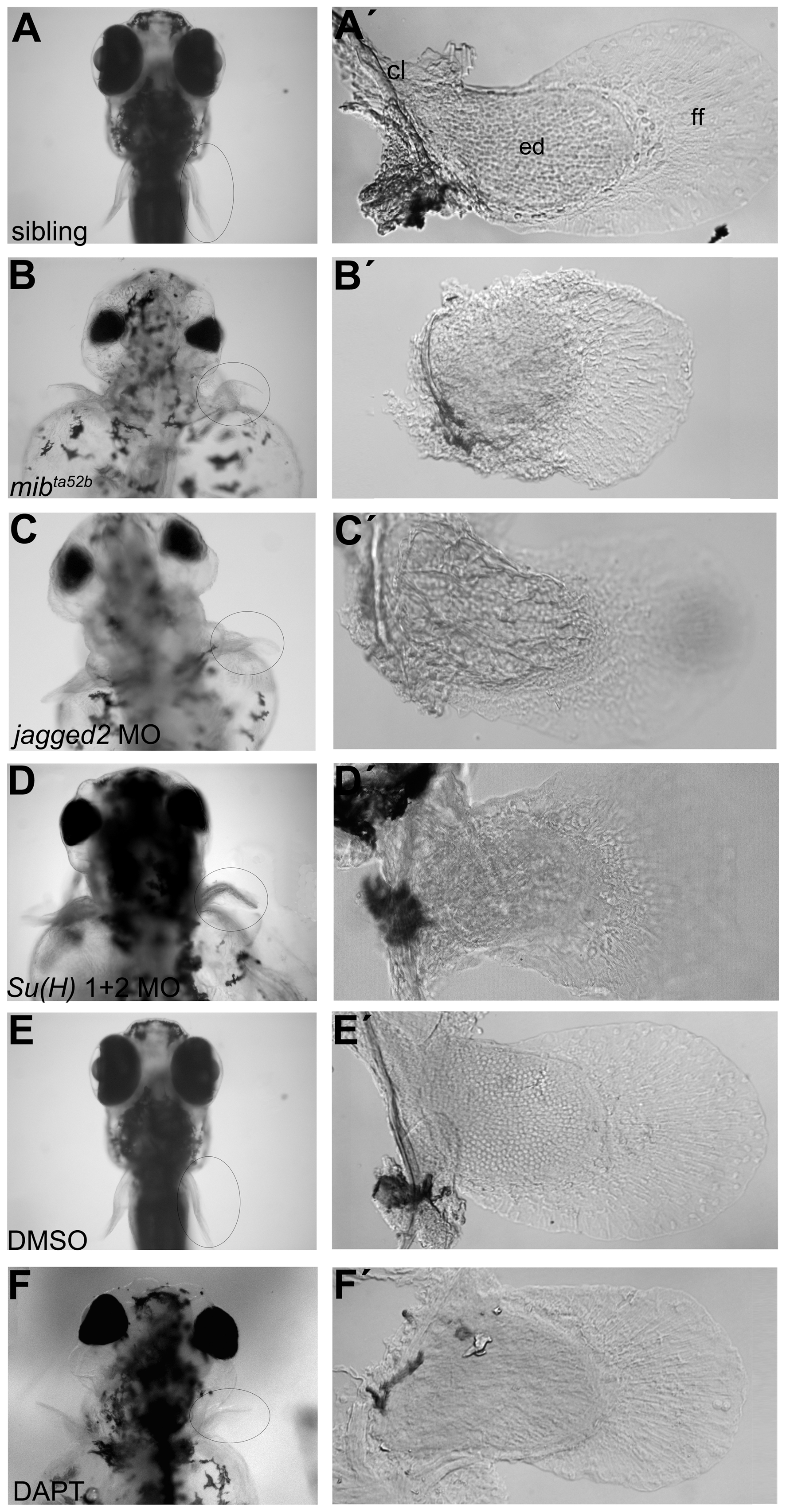
Fig. 2
Abnormal pectoral fins are formed in Notch signalling disrupted larvae.
(A-F) Live pictures of 5 dpf larvae. (A′) A pectoral fin from a sibling embryo showing the cartilaginous endoskeletal disc with individualized cells surrounded by thin matrix deposits, the fin fold and the chleitrum (n = 17) (E, E′). A similar pectoral fin was found in a DMSO-treated embryo (n = 9). Pectoral fins of Notch signalling disrupted embryos such as mibta52b (n = 18) (B, B′), jagged2 (n = 10) (C, C′) and Su(H)1+2 (n = 12) (D, D′) morphants and DAPT-treated embryos (n = 10) (F, F′) showing disorganized endoskeletal disc cells. cl, chleitrum; ed, endoskeletal disc; ff, fin fold.