
|
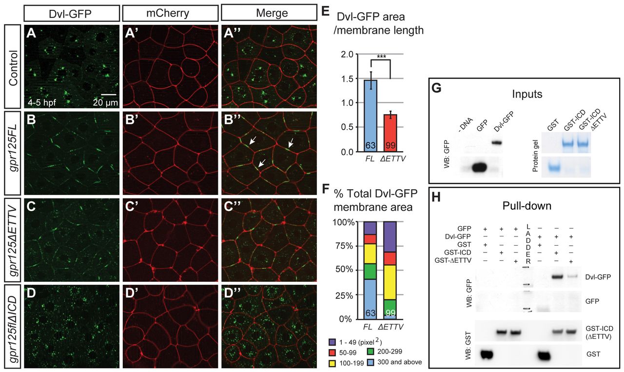
Fig. 5 Gpr125 promotes Dvl-GFP localization in discrete membrane subdomains via direct interaction. (A-D′) Animal pole views of live embryos at 4-5 hpf co-injected with 150 pg dvl-GFP and 50 pg mCherry RNA in the absence (A-A′) and presence of 380 pg gpr125FL (B-B′), gpr125ΔETTV (C-C′) or gpr125ΔICD RNA (D-D′). Arrows in B′ indicate Dvl-GFP membrane subdomains. (E) Ratio of Dvl-GFP membrane area to the length of the membrane measured on embryos expressing full-length or ΔETTV Gpr125. Numbers of membranes analyzed are inside bars. Data are mean±s.e.m. ***P<0.001. (F) Size distribution of Dvl-GFP membrane subdomains in embryos expressing full-length or ΔETTV Gpr125. Numbers of membranes analyzed are inside bars. (G,H) Pull-down assay with GST- and GFP-fusion proteins. Ten percent of GFP-fusion protein inputs were blotted with anti-GFP antibody and 100% GST fusion protein inputs were stained with Denville Blue Protein Stain (G). Pull-down results were analyzed by western blotting using anti-GFP and anti-GST antibodies (H).