
|
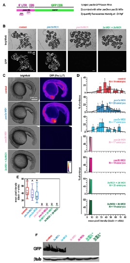
Fig. S9 pax3b, but not pax3a, MOs efficiently reduce translation of chimeric 5′UTRpax3b:GFP RNA. (A) Primers (forward with XhoI site 5′-gcgctcgagtgatcgtggaatattacatg-3; reverse with BamHI site 5′-gcgggatcc gagagtgatgggcgcatcat-3′) were used to amplify 62 bp of pax3b 5′UTR and 48 bp of pax3b-coding sequence. The resulting fragment was cloned in frame with EGFP in pEGFP-N1 (Invitrogen) to produce a 5′UTRpax3b:eGFP fusion protein. 5′UTRpax3b:eGFP was then subcloned into pSP64T, and RNA was transcribed using SP6 (Ambion). RNA (250 pg) was injected alone, or together with MO, into one-cell stage embryos. (B) At 24 hpf, embryos of control (5′UTRpax3b:eGFP) and 5′UTRpax3b:eGFP plus pax3a MO1 or MO2 contained high levels of GFP fluorescence. Embryos with either pax3b MO had no detectable fluorescence, indicating complete inhibition of 5′UTRpax3b:eGFP translation by pax3b MOs. (C) Images of single embryos were taken at identical magnifications and exposure. Background was normalized using the ‘Subtract Background’ feature of ImageJ (1.47n5). Pixel intensity of 8-bit greyscale images was visualized using the ‘Fire’ LUT. (D) Mean pixel intensity per embryo was normally distributed. (E) The mean pixel intensities were significantly reduced after addition of pax3b MO, as determined by ANOVA and Tukey’s post hoc test. (F) Protein lysates were separated by 12% SDS-PAGE and probed using antibodies against GFP or β-tubulin (loading control).