
Fig. S12
- ID
- ZDB-IMAGE-130816-4
- Publication
- Minchin et al., 2013 - Oesophageal and sternohyal muscle fibres are novel Pax3-dependent migratory somite derivatives essential for ingestion
- All Figures
- Figures for Minchin et al., 2013
|
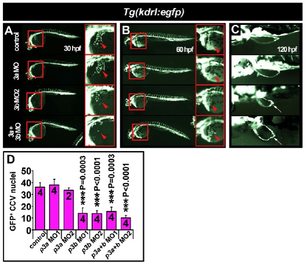
Fig. S12 Common cardinal vein development is delayed at 30 hpf in pax3b morphants. Fluorescent images of live embryos expressing eGFP under the kdrl (formerly known as flk1 or vegfr2) promoter (A-C). Whole-mount lateral (A,B) or dorsal (C) views, anterior towards the left. (A) In 30 hpf embryos, general vasculogenesis was unimpaired after pax3a/b knockdown. However, the forming common cardinal vein (CCV) was reduced in pax3b single and double pax3a/b morphants (arrowheads). Boxed areas indicate regions magnified in the corresponding panels on the right. (B) In 60 hpf larvae, the general integrity of the vascular system was indistinguishable between control and pax3a/b morphants. Endothelial cells of the CCV had spread across the yolk (arrowheads). (C) Pectoral fin vasculature developed normally in pax3a, pax3b and pax3a+pax3b double morphants. The pectoral fin is malformed after pax3b manipulation (arrows). (D) Quantification of eGFP+ CCV nuclei at 30 hpf, mean±s.e.m. Sample number is indicated within each bar.