
|
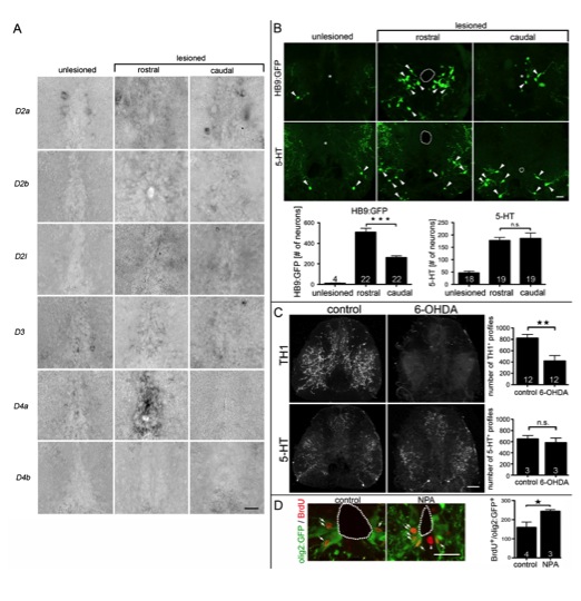
Fig. S6 (related to Fig. 7) Dopaminergic control of adult motor neuron regeneration. A: D4a, in contrast to other D2-like receptors, is strongly upregulated in the ependymal zone of the lesioned spinal cord only rostral to the lesion site at 14 days dpl. Spinal cross sections are shown (dorsal up). B: Small intensely labeled HB9:GFP+ motor neurons (arrowheads) are rarely found in unlesioned animals. After a lesion, significantly more (1.9x) HB9:GFP+ motor neurons are generated rostral than caudal to a spinal lesion site (***P < 0.0001). Numbers of 5-HT+ neurons (arrowheads) are strongly increased compared to those in unlesioned animals, but there is no rostrocaudal difference in number (P > 0.05). C: 6-OHDA destroys TH1+ axons, but not serotonergic (5-HT+) axons. Complete cross sections through the spinal cord are shown; dorsal is up. For control and 6-OHDA treatment, respectively, double immuno-fluorescent labeling of the same tissue section is shown. Whereas an intraperitoneal injection of 6-OHDA significantly (**P < 0.01) reduces the number of TH1+ axons, no effect on 5-HT+ axons was detectable (P > 0.05), indicating selectivity of the toxin. D: The number of BrdU+/olig2:GFP+ pMN-like cells (arrows) at the central canal (dotted line) was increased when motor neuron numbers were increased after NPA application (3, 6, 9 dpl) at 14 dpl (*P < 0.05, one-sided test). Error bars represent SEM. Scale bars: A: 15 μm; B: 50 μm; C: 40 μm; D: 50 μm.
Reprinted from Developmental Cell, 25(5), Reimer, M.M., Norris, A., Ohnmacht, J., Patani, R., Zhong, Z., Dias, T.B., Kuscha, V., Scott, A.L., Chen, Y.C., Rozov, S., Frazer, S.L., Wyatt, C., Higashijima, S., Patton, E.E., Panula, P., Chandran, S., Becker, T., and Becker, C.G., Dopamine from the Brain Promotes Spinal Motor Neuron Generation during Development and Adult Regeneration, 478-491, Copyright (2013) with permission from Elsevier. Full text @ Dev. Cell