
|
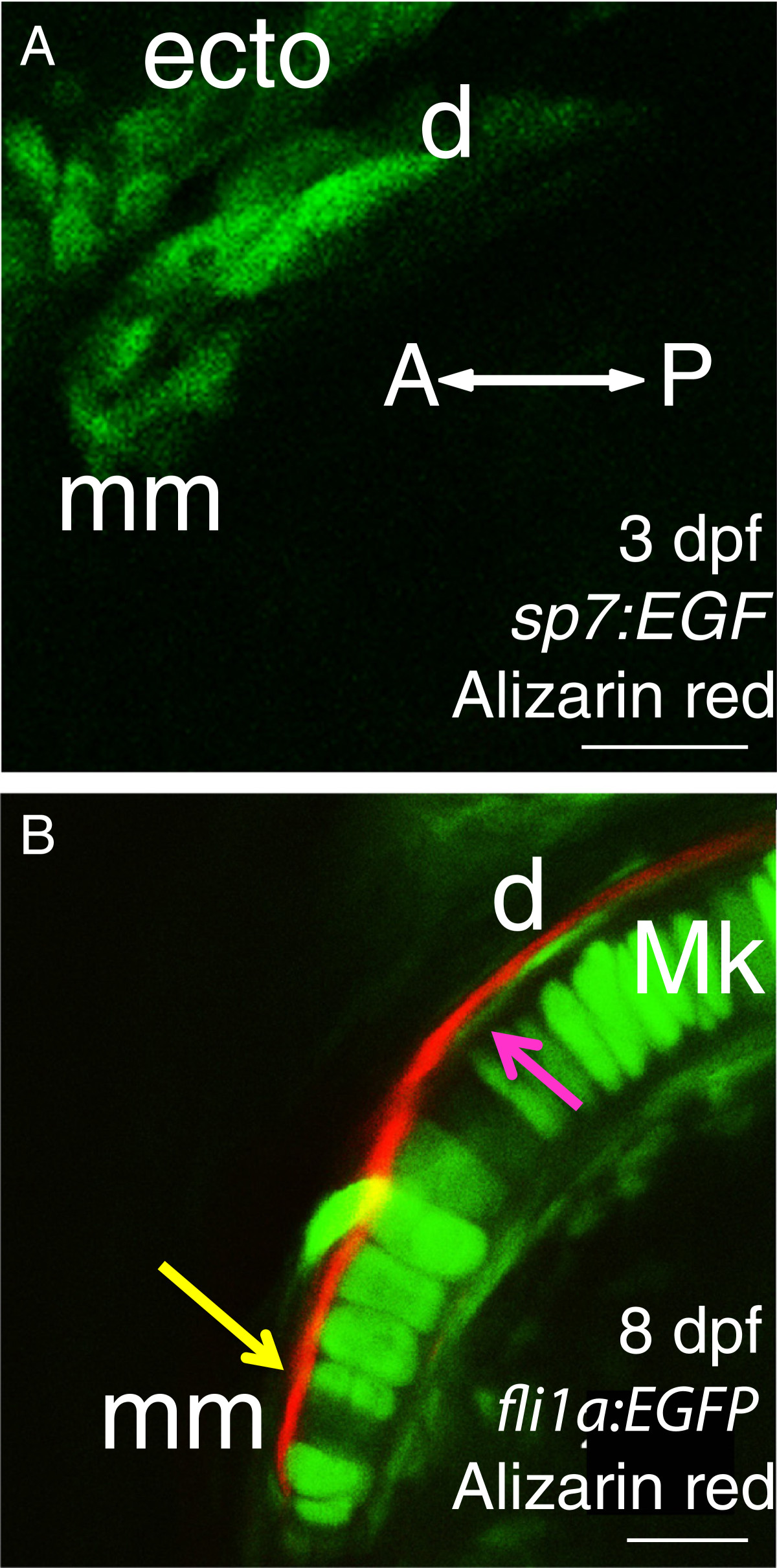
Fig. 5 Cellular resolution of bones in the anterior Meckel′s cartilage suggests that the dentary has fused with the mentomeckelian bone from the earliest stages of osteogenesis. Before bone matrix is detected by Alizarin red, only one group of osteoblasts is apparent adjacent to Meckel’s cartilage in 3 dpf Alizarin red-stained sp7:EGFP larvae from a ventral view (A). In a confocal slice of the anterior region of Meckel’s cartilage in 8 dpf Alizarin red-stained fli1a: EGFP larvae (B), a single bone is visible from a ventral view, but its association with chondrocytes of Meckel’s cartilage appears to vary anteroposteriorly. Anteriorly, bone lies immediately adjacent to chondrocytes (yellow arrow), while more posteriorly, the bone appears to be separated from chondrocytes by cells of the presumptive perichondrium (magenta arrow). Abbreviations: A = anterior; d = dentary; dpf = days post-fertilization; ecto = ectoderm; Mk = Meckel’s cartilage; mm = mentomeckelian; P = posterior. Scale bars: A,B = 20 μm.