
Image
|
Figure Caption
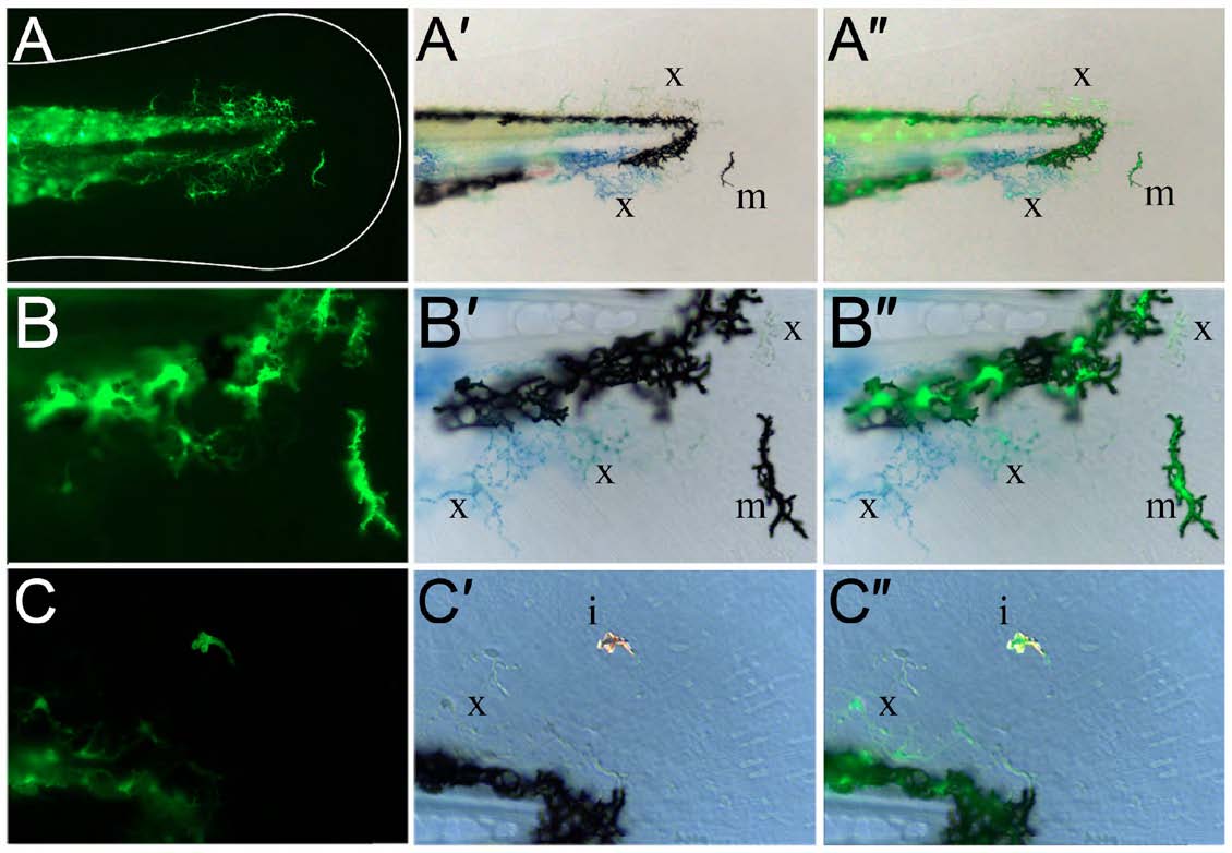
Fig. S2
Kaede-positive cells in the fin are chromatophores. (A-C") Lateral views of the fin region of three 3-dpf sox10:gal4; uas:kaede transgenic embryos displaying Kaede green fluorescence (A,B,C) and brightfield views (A′,B′,C′). An overlay of the fluorescence image on the brightfield image is also shown (A",B",C"). At both low (A-A") and high (B-C") magnification, it can be observed that the Kaede-positive cells in the fin (A,B,C) correspond to black melanophores (m; A′,A",B′,B"), xanthophores with characteristic yellow/blue colouration (x; A′,A",B′,B",C′,C") or reflective iridophores (i; C′,C").
Acknowledgments
This image is the copyrighted work of the attributed author or publisher, and
ZFIN has permission only to display this image to its users.
Additional permissions should be obtained from the applicable author or publisher of the image.
Full text @ Development