
Image
|
Figure Caption
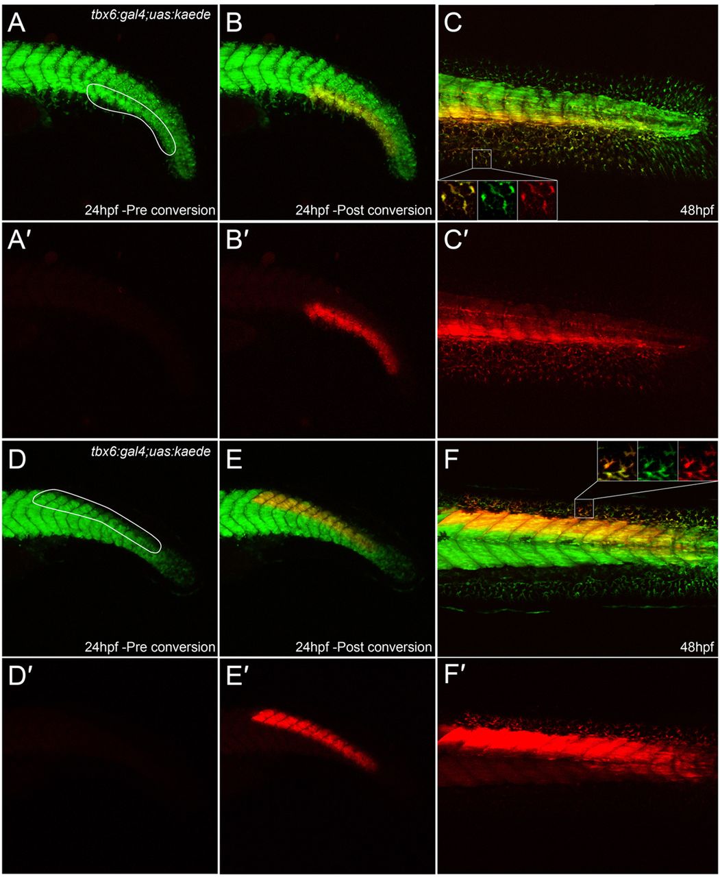
Fig. 3
Extensive contribution of mesoderm to fin mesenchyme. (A-F2) Tail of tbx6:gal4; uas:kaede embryos at 24 hpf (A-B2,D-E2) and 48 hpf (C,C2,F,F2) both prior to (A,A2,D,D2) and after (B-C2,E-F2) Kaede photoconversion. Unconverted Kaede protein is in green, overlaid with UV-photoconverted Kaede in red (A-F); the red channel is additionally displayed alone for clarity (A2-F2). Ventral (A-C2) and dorsal (D-F2) regions converted by UV laser are outlined in A and D. At 48 hpf, converted cells can be seen in the adjacent fins (magnified in insets in each channel and merged, C,F) and muscle blocks (C,C2,F,F2).
Figure Data
Acknowledgments
This image is the copyrighted work of the attributed author or publisher, and
ZFIN has permission only to display this image to its users.
Additional permissions should be obtained from the applicable author or publisher of the image.
Full text @ Development