
|
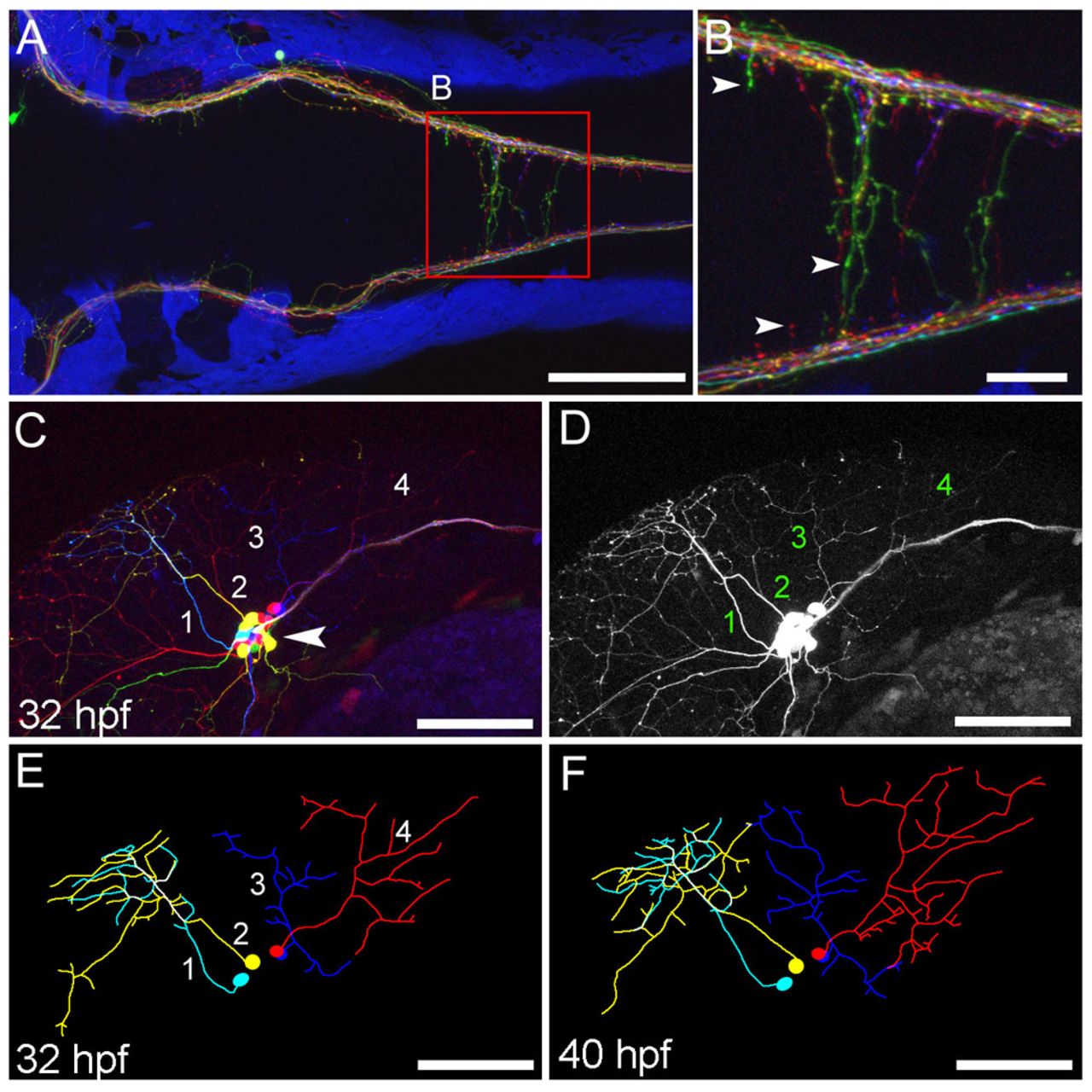
Fig. 4 Axon labeling and tracing. The s1102t;UAS:Zebrabow-V transgenic line labels somatosensory neurons and their axons. (A,B) Central axons in the hindbrain at 5 dpf, viewed dorsally. Axonal varicosities (presynaptic terminals) are visible in individual axons (B, arrowheads). (C-F) Somatosensory neuron cell bodies are located in the trigeminal ganglion (C, arrowhead). Each neuron forms an axonal arbor that branches extensively in the skin. Four neurons (numbered) were traced and are shown in E. The same image in the absence of color information is more difficult to trace (D). Axonal morphology was imaged every 2 hours from 28 to 44 hpf. Two time points are shown (E,F). Scale bars: 100 μm in A,C-F; 25 μm in B.