
|
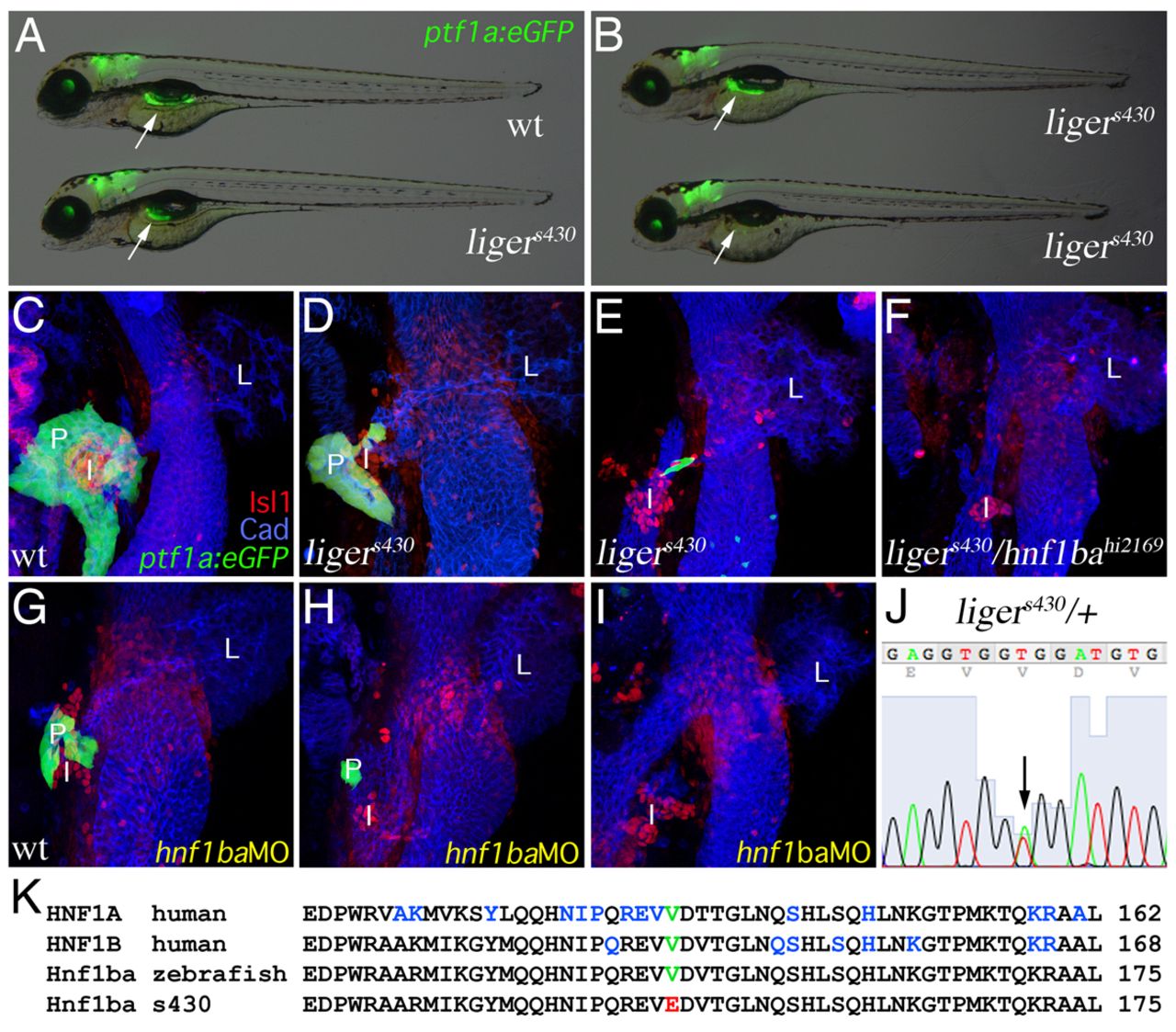
Fig. 1 ligers430 is a hypomorphic hnf1ba zebrafish mutant and exhibits MODY5-like pancreas hypoplasia. (A,B) Merged fluorescent/bright-field micrographs of 4 dpf Tg(ptf1a:eGFP)jh1 (arrows indicate pancreas) wild type (A, top) and ligers430 mutants showing the most common severity of pancreas hypoplasia (A, bottom). In ligers430 mutant embryos, the pancreas can be either nearly normal in size (B, top; <10%) or completely absent (B, bottom; <10%). (C-I) Three-dimensional rendering of the foregut endoderm of 80 hpf Tg(ptf1a:eGFP)jh1 wild-type (C), ligers430 (D,E), ligers430/hnf1bahi2169 (F) and hnf1ba-MO injected (G-I) embryos stained for Isl1 and cadherin demonstrating complementation failure by hnf1bahi2169 (F) and phenocopy with Hnf1ba translational knock-down (compare D-F with G-I, respectively). (J) Genomic sequence from a ligers430 heterozygote indicating a molecular lesion (arrow; double peek) in hnf1ba. (K) Amino acid alignment covering part of the atypical POU-specific domain of human HNF1A and HNF1B and wild-type zebrafish and ligers430 Hnf1ba. Red font denotes the valine to glutamic acid substitution at position 147 in ligers430. Blue font indicates amino acids affected by mis-sense mutations in MODY3 and MODY5.