
|
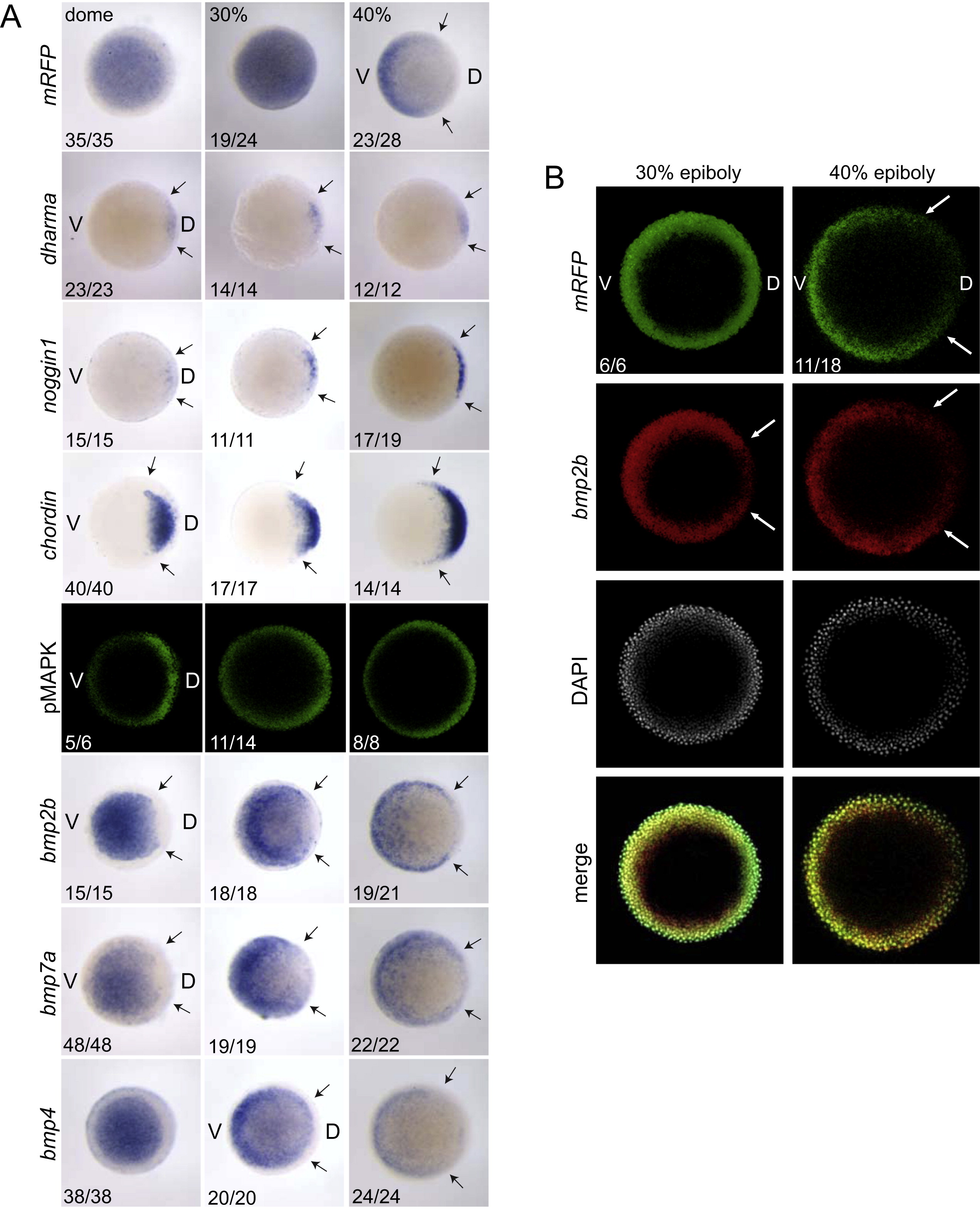
Fig. 4 The establishment of the BMP activity gradient occurs in a pre-established gradient of BMP ligand expression. (A) BRE-mRFP or wild-type embryos stained for mRFP, dharma, noggin1, chordin, bmp2b, bmp7a or bmp4 mRNA by ISH or immunostained for pMAPK. Arrows indicate the extent of the domain of staining for a particular mRNA. The number of embryos out of the total analysed that showed the presented staining pattern is given. (B) DFISH for mRFP and bmp2b in blastula stages BRE-mRFP embryos. The fluorescent substrates used were fluorescein tyramide (mRFP) and Fast Blue (bmp2b, pseudocoloured in red). The images are the projection of a 10–12 picture stack taken at the margin of the blastoderm to avoid fluorescence from the yolk. V, ventral; D, dorsal.
Reprinted from Developmental Biology, 378(2), Ramel, M.C., and Hill, C.S., The ventral to dorsal BMP activity gradient in the early zebrafish embryo is determined by graded expression of BMP ligands, 170-82, Copyright (2013) with permission from Elsevier. Full text @ Dev. Biol.