
Fig. 6
- ID
- ZDB-IMAGE-130801-32
- Genes
- Publication
- Wang et al., 2013 - miR-34b regulates multiciliogenesis during organ formation in zebrafish
- All Figures
- Figures for Wang et al., 2013
|
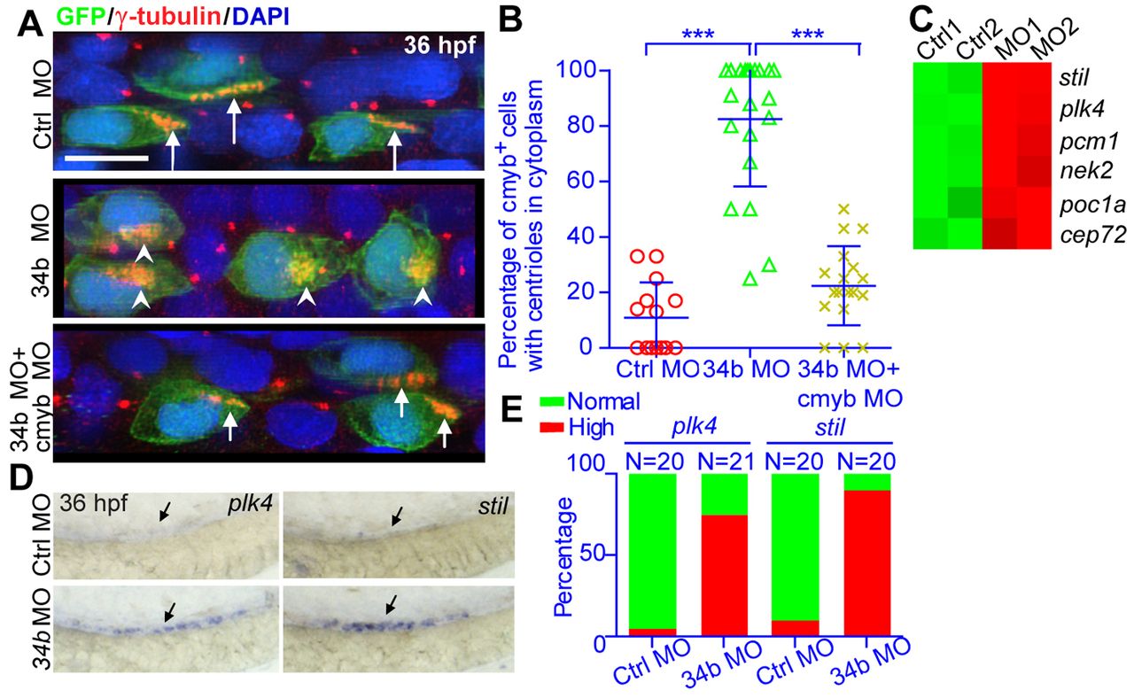
Fig. 6 The miR-34b-cmyb genetic pathway regulates the multiplication and membrane docking of centrioles. (A) The centriole (anti-γ-tubulin staining, red, arrows) morphology of MCCs (anti-GFP staining, green) in normal embryos, miR-34b morphants and embryos injected with both miR-34b MO and cmyb MO. Arrows indicate centrioles on the cell surface; arrowheads indicate centrioles in the cytoplasm. (B) Statistical analysis of A. Each circle/triangle/cross represents an embryo. Mean ± s.d. ***P<0.0001 (unpaired t-test). (C) Heat map showing the expression of genes related to centriole duplication from the microarray data of 3-dpf MCCs. The color scale represents the expression level of a gene above (red) or below (green) the mean expression level across all samples. (D) WISH analysis of plk4 and stil expression at 36 hpf. The arrow identifies the appropriate MCC region. (E) Statistical analysis of D showing normal versus high expression of plk4 and stil.