
Fig. 3
- ID
- ZDB-IMAGE-130801-19
- Genes
- Publication
- Nichols et al., 2013 - barx1 represses joints and promotes cartilage in the craniofacial skeleton
- All Figures
- Figures for Nichols et al., 2013
|
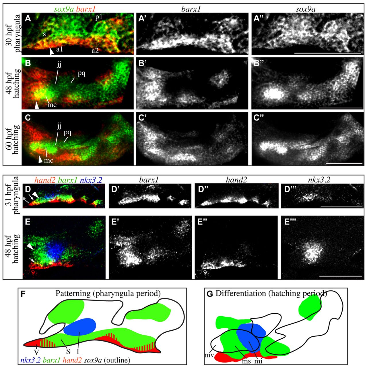
Fig. 3 barx1 is excluded from differentiating joint cells but maintained in the subintermediate domain. (A-C′) In situ hybridization was carried out to reveal wild-type barx1 expression (red) in the context of sox9a expression (green). (D-E′′) Fluorescent in situ hybridization revealed wild-type barx1 expression (green) in the context of hand2 (red) and nkx3.2 (blue) expression. Labeled pharyngula period anatomy includes the stomodeum (s), pharyngeal arch 1 (a1), pharyngeal pouch 1 (p1) and pharyngeal arch 2 (a2). Labeled hatching period anatomy includes the Meckel’s (mc) and palatoquadrate cartilages (pq), which are divided by the jaw joint (jj). Arrowheads indicate barx1 expression distal to the jaw joint in the subintermediate domain. Arrows indicate overlap between barx1 and hand2. Asterisk in D marks the ventral-most aspect of hand2 expression. In all images, anterior is towards the left, dorsal is upwards. All images are single confocal sections. Scale bars: 100 μm. (F,G) Outlines of gene expression data in A-E′′. Intermediate (I), subintermediate (S) and ventral (V) domains, and Meckel’s cartilage intermediate (mi), subintermediate (ms) and ventral (mv) are indicated.