
Fig. 1
- ID
- ZDB-IMAGE-130801-17
- Genes
- Publication
- Nichols et al., 2013 - barx1 represses joints and promotes cartilage in the craniofacial skeleton
- All Figures
- Figures for Nichols et al., 2013
|
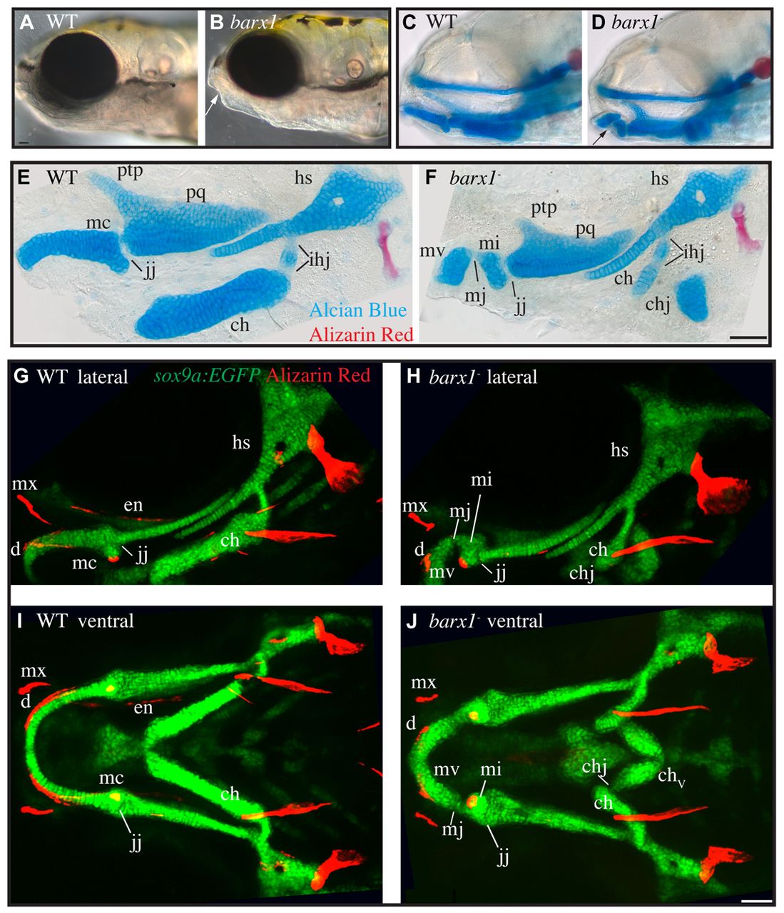
Fig. 1 Cartilage and dermal bone phenotypes present in barx1 mutant zebrafish. (A,B) Live 5 days post-fertilization (dpf) zebrafish larvae were imaged with transmitted light. Arrow indicates lower jaw divot. (C,D) 5 dpf zebrafish stained with Alcian Blue and Alizarin Red to label cartilage and bone were imaged with transmitted light. Eyes were removed to allow visualization of skeleton. Arrow indicates lower jaw skeletal gap. (E,F) 4 dpf Alcian Blue and Alizarin Red stained skeletons were dissected and imaged with transmitted light. (G-J) Confocal projections of live 6 dpf larvae with chondrocytes transgenically labeled with EGFP (sox9a:EGFP, green), and bone labeled with Alizarin Red. (A-H) Lateral views, anterior is towards the left, dorsal is upwards. (I,J) Ventral views, anterior is towards the left, whereas left is upwards. Arch 1 elements include the pterygoid process (ptp), palatoquadrate (pq) and Meckel’s (mc) cartilages. The jaw joint (j) and the ectopic Meckel’s joint (mj) are described in cellular detail below. The independent elements resulting from the ectopic arch 1 joint are referred to as Meckel’s cartilage ventral (mv) and Meckel’s cartilage intermediate (mi). Arch 1-derived dermal bones indicated are the dentary (d), maxilla (mx) and entopterygoid (en). Arch 2-derived cartilages include the ceratohyal (ch) and hyosymplectic (hs), which are divided by the interhyal joints (ihj) and the ectopic ceratohyal joint (chj). Scale bars: 50 μm.