
|
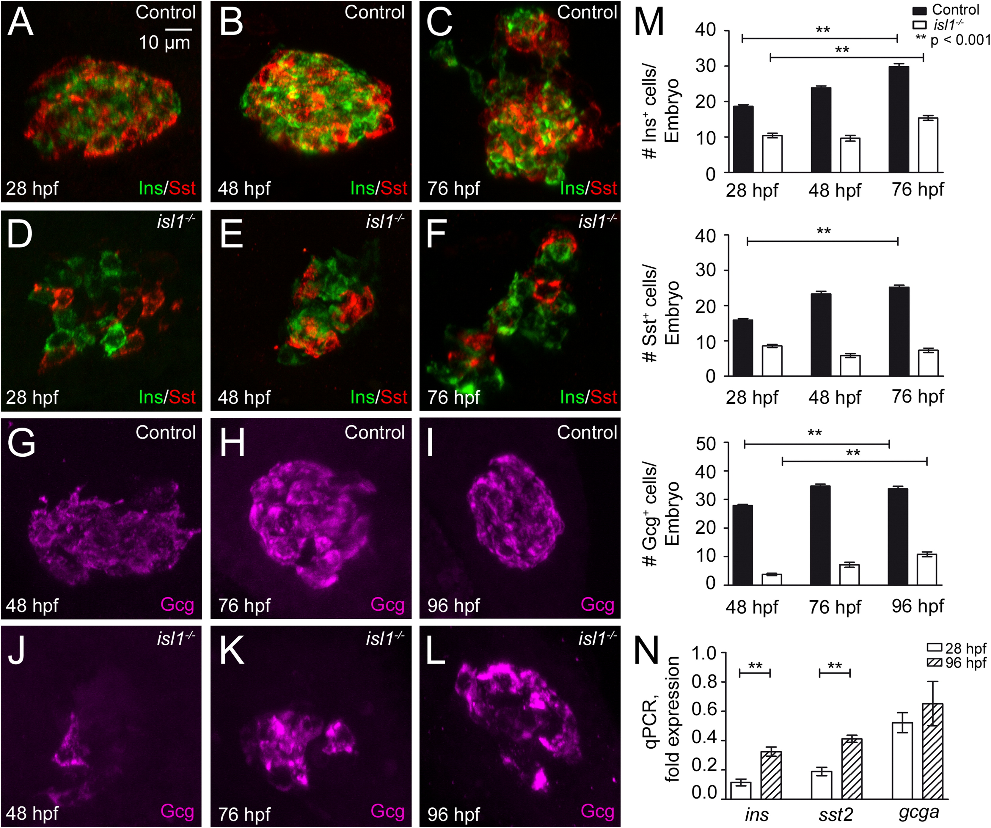
Fig. 1 Reduced expression of endocrine hormones in isl1 mutants. (A–F) Confocal image projections of the pancreatic islet in control (A–C) and isl1-/- embryos (D–F) that were co-immunostained for Ins (green) and Sst (red) at 28 hpf (A, D), 48 hpf (B, E) and 76 hpf (C, F). (G–L) Immunostainings for Gcg (purple) in control (G–I) and in isl1-/- embryos (J–L) at 48 hpf (G, J), 76 hpf (H, K) and 96 hpf (I, L). All embryos are shown from ventral with the anterior to the left. (M) Quantitative analysis of hormone expressing cells in control and isl1-/- embryos. Bars show mean+SEM. (N) Relative expression levels of ins, gcga and sst2 mRNA in isl1-/- mutant fish in relation to control littermates as revealed by qPCR analyses of whole embryo RNA preparations, normalized to EF1α.
Reprinted from Developmental Biology, 378(1), Wilfinger, A., Arkhipova, V., and Meyer, D., Cell type and tissue specific function of islet genes in zebrafish pancreas development, 25-37, Copyright (2013) with permission from Elsevier. Full text @ Dev. Biol.