
Fig. 2
|
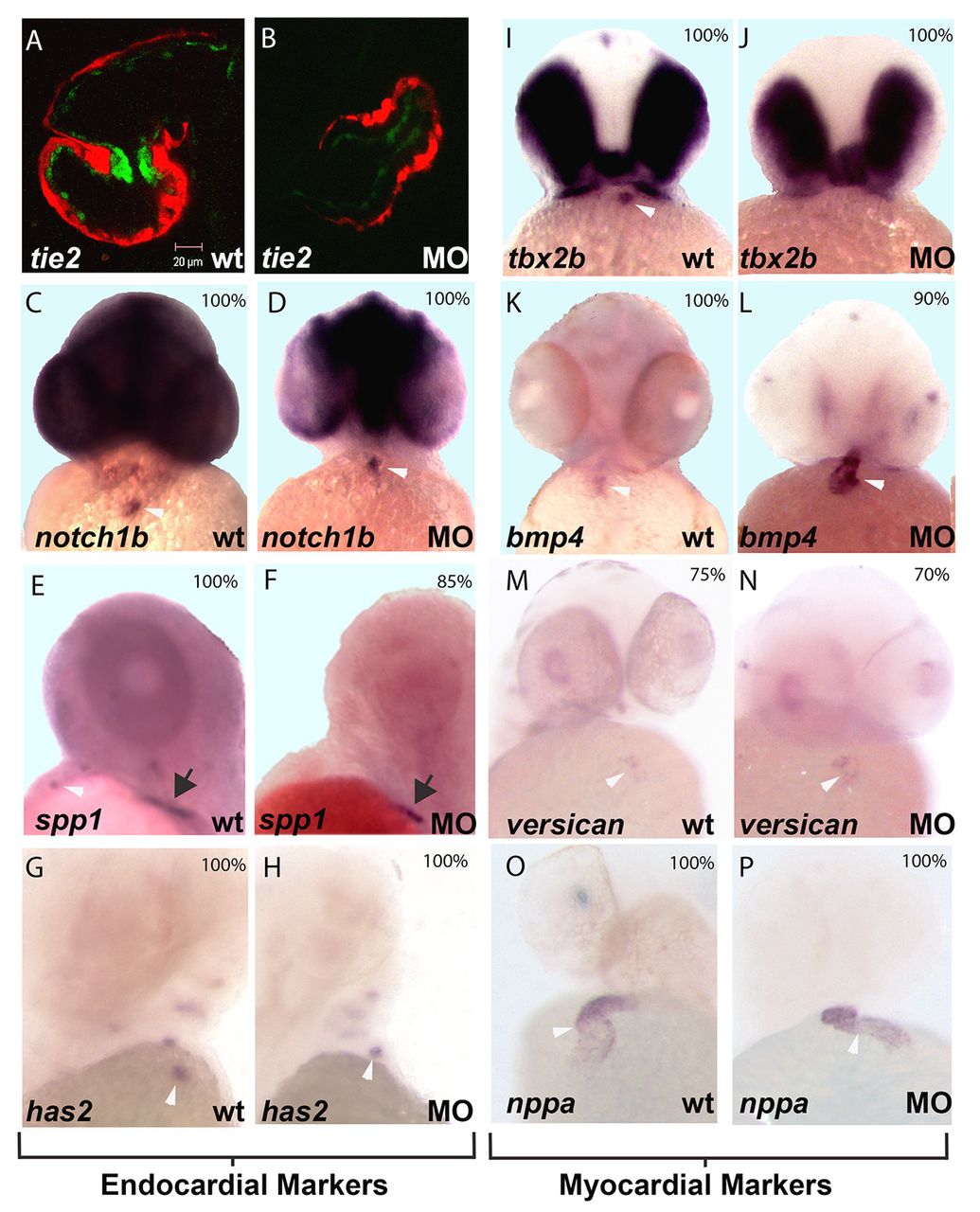
Fig. 2 miR-21 knockdown affects both endothelial and myocardial AV ring markers. (A,B) Confocal microscopy of control transgenic embryos (cMLC2:dsRed/Tie2:EGFP) show GFP expression (green) in pre-valve AV ring endothelium at 48 hpf (A), whereas miR-21 knockdown embryos do not (B). (C-P) In situ images are oriented with atrium at bottom and ventricle at top. The probe is indicated bottom left and the experimental condition [wild-type control (wt) versus miR-21 MO injected (MO)] bottom right. The percentage of embryos that display the representative in situ pattern is indicated top right (n=10-20). (C-H) Endocardial markers; (I-P) myocardial markers. White arrowheads indicate AV ring staining, where present. Black arrows (E,F) highlight spp1 expression in the fin bud. The majority of the AV ring markers are unchanged upon knockdown of miR-21. However, the myocardial marker tbx2b is absent from the AV ring after miR-21 knockdown (I,J), whereas bmp4 is expanded throughout the ventricle (L), as compared with control embryos (K).