
|
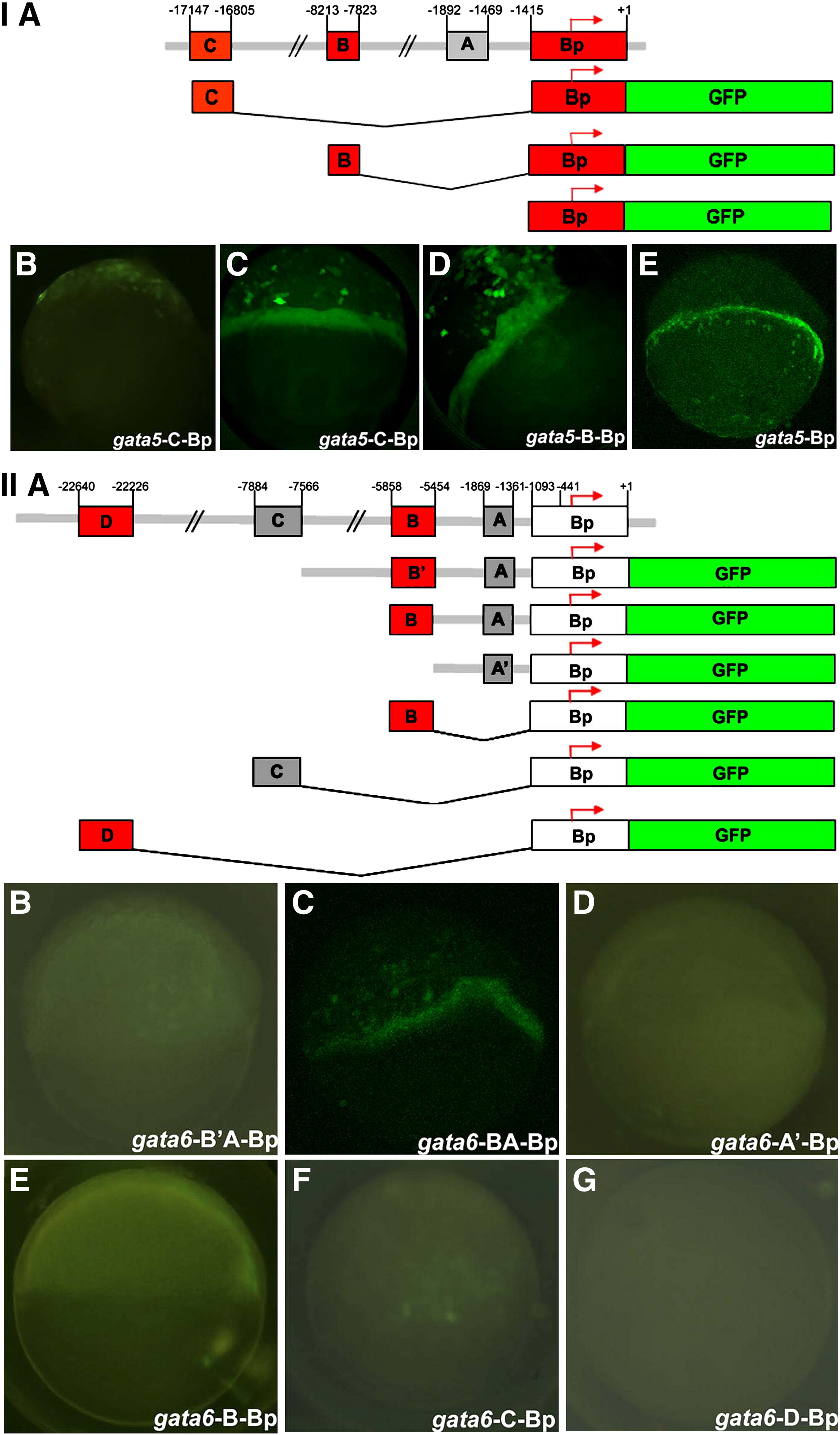
Fig. 8 Functional analysis of gata5 and gata6 regulatory modules. I. The GFP expression constructs, expression territories, and the GFP images of the gata5 regulatory module. (A) Map and summarization of the results for the gata5 regulatory modules. (B) gata5-C-Bp shows GFP in the dorsal half the embryo. (C) gata5-C-Bp shows GFP in the mesendoderm and scattered expression in the ectoderm. (D) gata5-B-Bp shows GFP in the mesendoderm and scattered expression in the ectoderm. (E) gata5-Bp shows GFP in the mesendoderm. II. GFP constructs and results for gata6 and representative images of the expression pattern. (A) Map and summarization of the results for the gata6 regulatory modules. (B) gata6-B2A-Bp shows GFP in the mesendoderm and YSL. (C) gata6-BA-Bp shows GFP in the mesendoderm. (D) gata6-A′-Bp shows no GFP expression in the embryo. (E) gata6-B-Bp shows GFP in the YSL. (F) gata6-C-Bp shows GFP in the YSL. (G) gata6-D-Bp shows no GFP expression in the embryo.
Reprinted from Developmental Biology, 357(2), Tseng, W.F., Jang, T.H., Huang, C.B., and Yuh, C.H., An evolutionarily conserved kernel of gata5, gata6, otx2 and prdm1a operates in the formation of endoderm in zebrafish, 541-57, Copyright (2011) with permission from Elsevier. Full text @ Dev. Biol.