
|
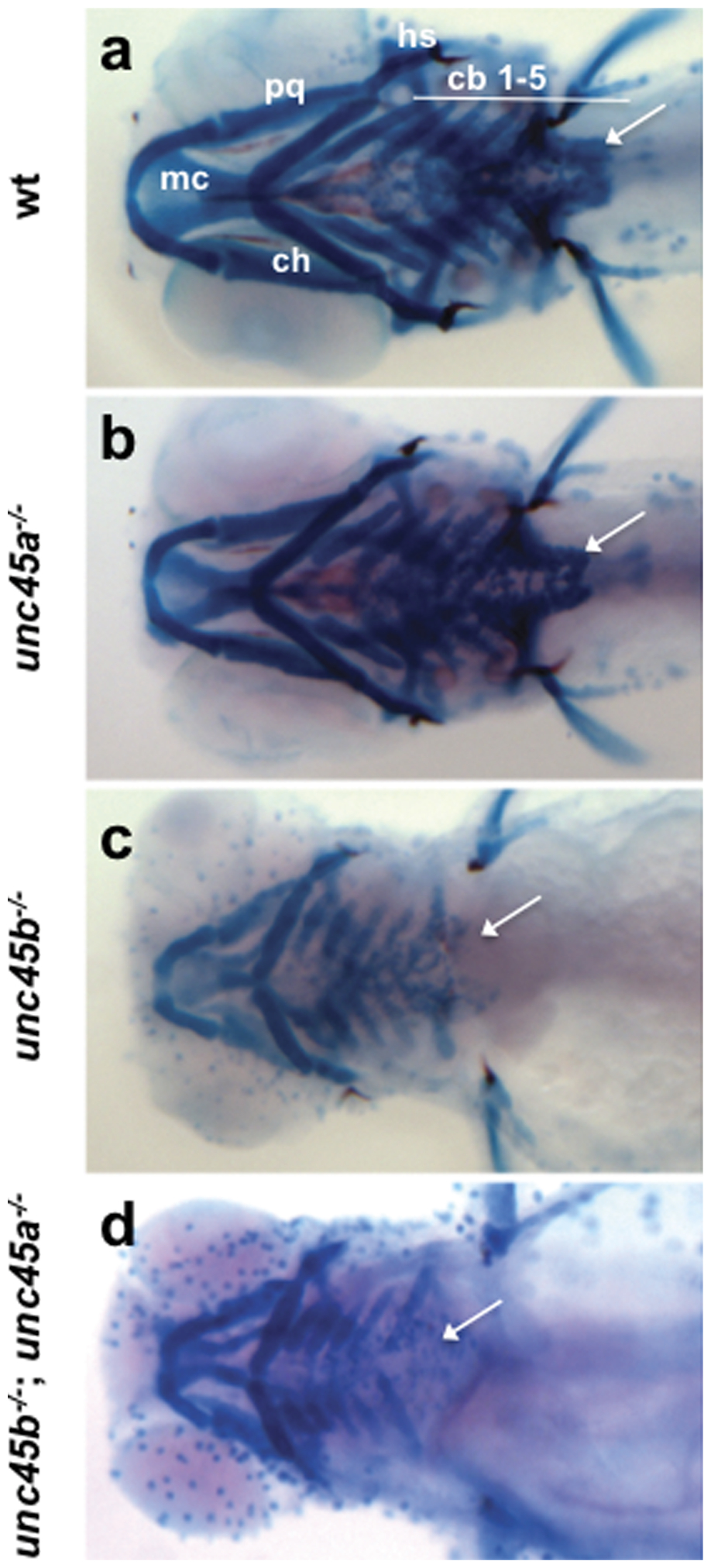
Fig. 6 Skeletal defects in unc45 mutants at 5 dpf.
Ventral views of Alcian Blue stained cartilages (a–d). Wild type siblings (a), unc45a-/- (b), unc45b-/- (c), and unc45b-/-; unc45a-/- (d) mutants. Wild type siblings and unc45a-/- mutants (a,b) have robust cartilage staining whereas unc45b-/- (c) and unc45b-/-; unc45a-/- (d) mutants exhibit decreased staining, improper angling of the ceratohyal cartilages, and shortening of the palatoquadrates and Meckel′s cartilage. The pectoral girdle (arrows) is missing or reduced in unc45b/ (c) and unc45b-/-; unc45a-/- (d) embryos. cb, ceratobranchial; ch, ceratohyal; hs, hyosymplectic; mc, Meckel′s cartilage; pq, palatoquadrate. Small blue dots are an artifact of the fixation and staining process.