
|
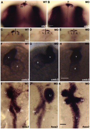
Fig. 4 Loss-of-function of dyx1c1 caused L–R assymetry defects in zebrafish.
Whole-mount in situ hybridization showing the expression of laterality markers in WT and dyx1c1 morphants (ATGMO+SPMO) at 2 dpf. The position of the asymmetrically placed organs was irregular in at least 50% of the embryos injected with dyx1c1 MO. otx5 expression showing the left sided placement of the parapineal organ in WT (A). In dyx1c1 morphant, the parapineal position was reversed (B). lov was strongly expressed in the left habenular in WT (C). In dyx1c1 morphant lov expression was stronger in the right or expressed symmetrically (D & E). Knockdown of dyx1c1 altered heart looping. Ventral views of embryos at 2 dpf (F–H). cmlc2 expression showed normal heart looping in WT (F). Cardiac looping was reversed (G) or absent in dyx1c1 morphant embryos (H). Expression of foxa3 revealing the position of the gut, liver and pancreas in WT embryo (I). The positions of the liver, gut and pancreas were irregular in dyx1c1 morphants (J & K). Panels A, B, C, D, E, I, J and K are dorsal views with anterior to the top. Frontal views are shown in panels F–G. Scale bars indicate 50 μm. Abbreviations: p, pineal organ: pp, parapineal organ: v, ventricle: a, atrium: li, liver: pa, pancreas: g, gut.