
|
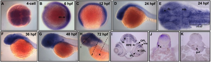
Fig. 4 Expression pattern of zebrafish PRL-1 during embryonic development. (A-D, and F-H) Whole mount in situ hybridization using probes against zebrafish PRL-1. Dorsal view of 4-cell stage (A) and 6-hpf (B) embryos. Lateral view of 12-hpf (C), 24-hpf (D), 36-hpf (F), 48-hpf (G), and 72-hpf (H) embryos. (E) Flat mount showed zebrafish PRL-1 signals in the entire brain. (I-K) Cross-sections along the plane indicated by lines showed in (H). Abbreviations: hpf, hours post fertilization; OPL, outer plexiform layer; OFL, optic fiber layer; IPL, inner plexiform layer; RPE, retinal pigment epithelium; dc, diencephalon; mb, midbrain; te, tectum; s, somite.