
|
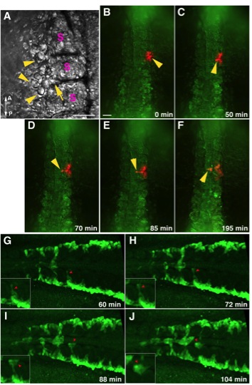
Fig. S2 The intersomitic migration of angioblasts in photoactivated Kaede mRNA injected embryos and Tg(fli1a:GFP) embryos. (A-F) Images extracted from time lapse video depict the intersomitic midline migration of photoconverted angioblasts. (A) Brightfield image before photoactivation showing angioblasts (arrowheads) clustering at the boundary of adjacent somites (S). Arrow depicts angioblasts ingressing into the somitic boundary. (B-F) In Kaede mRNA injected embryos, a cluster of cells that included anterior trunk angioblasts were photoconverted for differential labeling. Following photoconversion, the time progression shows activated angioblasts migrating between somites (C-E; arrowhead) towards the midline (F). Some cells in the somitic mesoderm were labeled as well during photoconversion. Scale bar for (A) and (B-F) represent 50 and 100 μm, respectively. A – anterior; P – posterior; S – somites. (G-J) Images extracted from time lapse confocal imaging of a dorsally mounted fli1a:GFP embryo showing angioblast and erythroid cell migration. Red arrows depict the intersomitic migration of angioblasts towards the midline. Insets show magnified views of migrating angioblasts.
Reprinted from Developmental Cell, 25(2), Kohli, V., Schumacher, J.A., Desai, S.P., Rehn, K., and Sumanas, S., Arterial and venous progenitors of the major axial vessels originate at distinct locations, 196-206, Copyright (2013) with permission from Elsevier. Full text @ Dev. Cell