
|
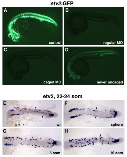
Fig. S4 The effects of photoactivatable etv2 MO on Tg(etv2:GFP) fluorescence and etv2 expression. (A-D) Photoactivatable etv2 MO inhibits etv2-2:GFP fluorescence. (A) Control uninjected, (B) regular etv2 MO2 injected embryo, (C) caged etv2 MO injected embryo uncaged at the sphere stage (4 hpf), (D) caged etv2 MO injected embryo never uncaged. All embryos were analyzed at 24 hpf with the same imaging and acquisition settings. (C) Embryos that were uncaged at the sphere stage resulted in nearly complete inhibition of GFP expression, showing a similar phenotype to standard etv2 MO injected embryos. (D) Caged MO injected embryos that were never uncaged showed higher GFP fluorescence than (C) uncaged embryos. While caged MO injected embryos that were never uncaged (D) showed a decrease in GFP expression in comparison to (A) uninjected embryos, these embryos were phenotypically normal with no apparent vascular defects. (E-H) etv2 expression analyzed in flat mounted embryos at the 22 to 24-somite stage in (E) control uninjected, (F) caged MO injected embryos that were uncaged at the sphere, (G) 5-somite and (H) 10-somite stage. Dorsal view, trunk and tail region is shown. (E) Medial (arrowhead) and lateral (arrows) angioblasts are shown migrating, and many cells have reached the midline. (F, G) Midline migration is absent in embryos that were uncaged at the sphere, and the 5-somite stage. (H) In embryos uncaged at the 10-somite stage, the medial angioblasts migrate to the midline while the migration of lateral angioblasts is inhibited.
Reprinted from Developmental Cell, 25(2), Kohli, V., Schumacher, J.A., Desai, S.P., Rehn, K., and Sumanas, S., Arterial and venous progenitors of the major axial vessels originate at distinct locations, 196-206, Copyright (2013) with permission from Elsevier. Full text @ Dev. Cell