
|
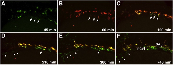
Fig. 5 Migration of Medial and Lateral Angioblasts to the DA and PCV Observed in Live etv2:Kaede Embryos Selected frames from a time-lapse movie of a single etv2:Kaede embryo prephotoconversion at the 14-somite stage (A) and postphotoconversion starting at the 15-somite stage (B) through 28 hpf (F). Arrows mark laterally positioned angioblasts that migrate directly to a ventral position to contribute to the PCV. Arrowheads mark a separate population of lateral angioblasts that contribute directly to the common cardinal vein (CCV). All views are dorsolateral; anterior is to the left, and dorsal is to the top. All selected frames are from Movie 6, embryo 1; imaging (t = 0 min) was started at approximately 13-somite stage. See also Movies S5, S6, S7, and S8.
Reprinted from Developmental Cell, 25(2), Kohli, V., Schumacher, J.A., Desai, S.P., Rehn, K., and Sumanas, S., Arterial and venous progenitors of the major axial vessels originate at distinct locations, 196-206, Copyright (2013) with permission from Elsevier. Full text @ Dev. Cell