
|
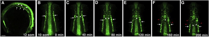
Fig. 3 The Migration of Trunk Angioblasts in Live etv2:GFP-Transgenic Embryos(A–G) GFP expression is observed in the medial and lateral endothelial progenitor cells at the 10- to 16-somite stages. In laterally (A) and dorsally mounted embryos (B–G), medial angioblasts (white arrows) migrate toward the midline (white arrowheads) intersomitically. (B–G) Selected frames from a time-lapse movie showing migration of the medial angioblasts; imaging was started at the 10-somite stage. The anterior angioblasts migrate first, followed by the more posterior endothelial progenitor cells. (F and G) Faint GFP expression in the lateral angioblasts (red arrowheads) becomes apparent; these cells migrate to the midline similar to the medial angioblasts. In (A) anterior is to the left; in (B)–(G) anterior is up. Scale bar, 100 μm. See also Figure S2 and Movies S1, S2, S3, and S4.
Reprinted from Developmental Cell, 25(2), Kohli, V., Schumacher, J.A., Desai, S.P., Rehn, K., and Sumanas, S., Arterial and venous progenitors of the major axial vessels originate at distinct locations, 196-206, Copyright (2013) with permission from Elsevier. Full text @ Dev. Cell