
|
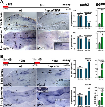
Fig. 3 Heat-shock–induced manipulation of Hedgehog (Hh) signaling at larval and juvenile stages. A: Reduced ptch2 expression in the ventricular regions of the brain 6 hr post heat shock in hsp:gli2DR transgenic 1-week-old larvae (compare top two panels) and 1-month-old juveniles (bottom two panels), as visualized by in in situ hybridization (ISH). Quantitative polymerase-chain reaction (qPCR) analysis revealed that ptch2 mRNA levels were reduced approximately 70% and 30% relative to non–heat-shocked 1-week and 1-month transgenics, respectively (right). EGFP mRNA levels were increased 4.8- and 2.2-fold in larvae and juveniles, respectively. B: Regionally increased Shh signaling in double heat-shocked hsp:shh transgenic fish visualized by ISH. In larvae, ptch2 expression was similar to non–heat-shocked transgenic and wild-type fish when examined by ISH, with ectopic expression seen in the anterior tectum (arrows). In juveniles, ptch2 expression was increased in its normal expression domain. No changes in ptch2 gene expression were seen in non–heat-shocked transgenic fish (right insets). EGFP mRNA was induced throughout the central nervous system in juveniles (inset). qPCR analysis revealed 1.8- and 1.6-fold increases in ptch2 expression in larvae and juveniles, respectively. EGFP mRNA levels were increased 15- and 25-fold, indicating effective heat-shock activation of the transgene at both ages. cb, cerebellum; di, diencephalon; hb, hindbrain; tec, tectum; tel, telencephalon. ***P < 0.001, **P < 0.01, *P < 0.05.