
|
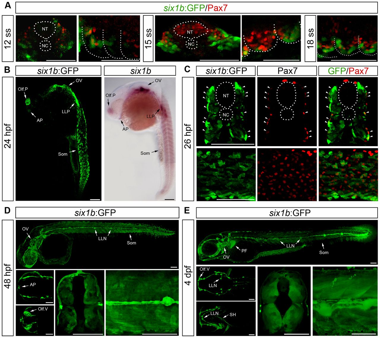
Fig. 3 Expression pattern of six1b:GFPJ1. (A) Expression of Pax7 (red) and GFP (green) in cross-sections at 12 and 15ss and lateral view of somite number 2–3 at 12, 15 and 18ss. (B) GFP expression of six1b:GFP at 24hpf phenocopying mRNA expression of six1b. (C) Cross-section at somite number 10 and lateral view of six1b:GFP (green) stained with Pax7 (red); arrowheads indicate coexpression. (D,E) GFP expression in lateral view of whole embryo; dorsal (top) and ventral (bottom) view of head; and cross-section at somite number 8 and enlargement of trunk of six1b:GFP embryos at 48hpf (D) and 4dpf (E). AP, adenohypophyseal placode; LLN, lateral line neuromasts; LLP, lateral line placode; NC, notochord; NT, neural tube; Olf.P, olfactory placode; Olf.V, olfactory vesicle; OV, otic vesicle; SH, sternohyoideus; Som, somites; PF, pectoral fin. Scale bars: 100μm.