
Image
|
Figure Caption
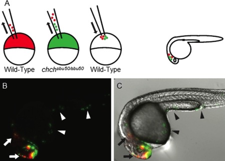
Fig. 8 churchill mutant cells exhibit increased spreading compared with wild-type. A: A schematic of the experimental procedure. wild-type and MZchchsbu50/sbu50 donors were labeled with rhodamine (red) and fluorescein (green) dextrans, respectively, and co-transplanted into unlabeled wild-type host embryos. Host embryos were observed at 24 hpf to determine the extent of cell spreading. B,C: A fluorescent only (B) and a merge of fluorescent and DIC images (C) indicate that chch mutant cells mobilize to posterior regions of the embryo (arrowheads), whereas wild-type cells remain in or close to the head (arrows).
Acknowledgments
This image is the copyrighted work of the attributed author or publisher, and
ZFIN has permission only to display this image to its users.
Additional permissions should be obtained from the applicable author or publisher of the image.
Full text @ Dev. Dyn.