
|
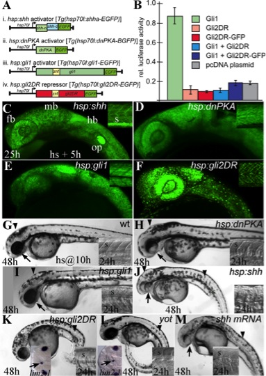
Fig. 1 Heat-shock–inducible activation and repression of Hedgehog (Hh) signaling. A: Four transgene constructs designed to allow heat-shock manipulation of Hh/Gli signaling. (i) hsp:shh activator line [Tg(hsp70l:shha-EGFP)]: (ii) hsp:dnPKA activator line [Tg(hsp70l:dnPKA-BGFP)]: (iii) hsp:gli1 activator line [Tg(hsp70l:gli1-EGFP)]. (iv) hsp:gli2DR repressor line [Tg(hsp70l:gli2DR-EGFP)]. B: Luciferase reporter assay. The modified Gli2DR protein and the Gli2DR-EGFP fusion protein caused an 80% reduction in luciferase activity in the presence of Gli1. C–F: Live images of 25 hours postfertilization (hpf) transgenic embryos, 4 hr after the completion of a 1-hr heat shock. Insets show green fluorescent protein (GFP) expression in the somites of the trunk. C: In the hsp:shh line, diffuse GFP expression is consistent with cytoplasmic localization of the Shhc-term-EGFP fusion protein. D,E: Cytoplasmic expression of the dnPKA-BGFP fusion and Gli1-EGFP fusion proteins. F: The Gli2DR-EGFP fusion protein is localized to nuclei. G–K: 48 hpf embryos that were heat shocked at 10 hpf. G: Wild-type embryo, inset shows V-shaped somites at 24 hpf. H: hsp:dnPKA embryo displaying Hh over expression defects including ventrally reduced eyes (arrow), dorsal brain defects (arrowhead), and slightly flattened somites at 24 hpf typical of Hh over-expressing embryos (Koudijs et al., 2008). I: hps:gli1 embryo with no visible Hh over-expression defects. Somites appear morphologically normal at 24 hpf (inset). J: hsp:shh embryo showing more severe Hh gain of function defects including flattened somites (arrowhead and inset) and reduced eyes (arrow). K: hsp:gli2DR embryo showing defects associated with a loss of Hh signaling, including U-shaped somites (arrowhead and right inset) and ventrally positioned but well formed eyes. lim2.1 labeling reveals an ectopic midline lens (arrow, left inset) in a 30 hpf hsp-gli2DR embryo that was heat shocked at the 8-somite stage. L: Forty-eight hpf yot(gli2DR) mutant embryo with Hh loss of function defects including u-shaped somites (arrowhead and right inset) and a lim2.1-labeled ectopic midline lens (left inset, arrow). M: Hh overexpression defects in the somites (arrowhead) and forebrain (arrow) caused by injection of 100 pg of shh mRNA at the 2-cell stage. fb, forebrain; hb, hindbrain; mb, midbrain; op, otic placode; s, somite.