
|
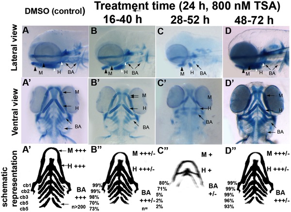
Fig. 5 Differential temporal requirements for HDAC function during craniofacial development.
A–D and A′–D′ Wild-type embryos treated with TSA for 24 hpf at different stages of embryonic development. After treatment periods other than 48–72hpf, embryos were washed to remove TSA and then allowed to develop until 3.5 dpf. Embryos were fixed at 3.5 dpf and then stained with alcian blue. A–D lateral views, A′-D′ ventral views. A–A′ are DMSO-treated controls, B–B′ 16–40 hpf TSA-treated embryos, C–C′ 28–52 hpf TSA-treated embryos, D-D′ 48–72 hpf TSA-treated embryos. A′′-D′′ schematic with summary of craniofacial defects at different TSA treatment concentrations. M, mandibular; H, hyoid; cb1-5, cerato-branchials 1-5; BA, branchial arches;+++ wild-type,++ reduced in size compared to wild-type,+Severely reduced compared to wild-type, +/ severely reduced or absent altogether.