
|
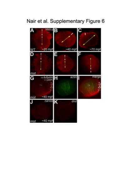
Fig. S6 The cortical band of germ plasm RNPs fails to undergo a peripherally-directed compression in motley/birc5b mutants. Animal views of blastodisc cortex, immunolabeled with anti-NMII-p antibody. (A–F) Segregation of GP RNPs during the first two cycles. In wild-type embryos at 20mpf, the center of the blastodisc cortex is GP RNP-free as the RNPs are located in a broad peripheral band (A). As development proceeds, the blastodisc center remains free of GP RNPs and the peripheral band becomes compressed from the center outwards as the central GP RNP-free zone expands (B, C). In motley/birc5b mutants at 20mpf, the center of the blastodisc is GP RNP free and the RNPs are in a broad peripheral band as in wild-type embryos (D). However, the peripheral GP RNP band fails to further compress noticeably during development (E, F). Yellow double-headed arrows represent GP RNP-free zone in the blastodisc center, which does not expand in motley mutants. White double-headed arrows represent peripheral compression of GP RNP band in wild-type embryos, which does not occur effectively in motley/birc5b. (G–I) In motley mutants at a stage coincident with observed GP RNP segregation defects, F-actin (H,I) forms large bundles randomly crisscrossing the blastodisc, instead of circumferential bundles as observed in wild-type (Figure 5B, 5C, 5H; [16]). Such randomly oriented bundles also form in embryos after inhibition of microtubule polymerization [16] and may be related to F-actin gelation observed in early embryonic extracts [55]. DAPI labeling in (G,I) shows center of blastodisc; astral microtubules in G are not clearly visible as this time point coincides with the cyclical disassembly of this structure after reaching the cortex [16], [56]. (J,K) In situ hybridizations to detect germ plasm RNAs nanos (J) and dead end (K) in motley mutant embryos, showing defects in germ plasm segregation similar to those observed when visualizing GP RNPs with anti-NMII-p antibodies (compare to wild-type in Figure S5D, S5E; GP RNP recruitment in wild-type furrows can be detected as early as 30 mpf [16]).