
|
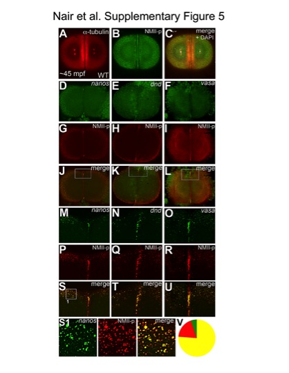
Fig. S5 Germ plasm mRNPs are labeled by anti-human phosphorylated non-muscle myosin II antibody. Animal views of 2-cell stage embryos; immunolabelings (A–C), fluorescent in situ hybridizations for nanos (D, M), dnd (E, N) and vasa (F, O), combined with immunolabeling for NMII-p (G–I, P–R). (M–U) Higher magnifications of boxed areas in (J–L). NMII-p labeling recapitulates the known patterns of germ plasm mRNA localization to the distal furrows and peripheral cortex in wild-type embryos at ~45mpf (A–C). nanos, dnd and vasa mRNAs label germ plasm aggregates at the cortical periphery and at the distal ends of the cleavage furrow (D–F, M–O). NMII-p expression overlaps with nanos (J, S), dnd (K, T) and vasa (L, U) both at the outlying cortex and the distal cleavage furrow. (S1) Higher magnification view of an example panel used for the quantitation of colocalization shown in (V), from the image in (S). (V) Pooled counts of observed particles (singletons and multimerized, n = 349) indicate that 76% of particles show both NMII-p and GP RNA labeling (yellow), 19% exhibit only NMII-p labeling (red) and 5% only GP RNA labeling (green). Due to the significantly higher sensitivity of the anti-NMII-p immunofluorescence labeling in comparison to the FISH technique to detect GP RNAs, it is likely that a significant fraction, if not most, of the 19% of particles that exhibit only NMII-p labeling also contain undetected GP RNAs. While we can not rule out that at these stages a minority of particles contain NMII-p without GP RNAs, the observation that less than 5% of particles apparently containing GP RNAs are not labeled with the anti-NMII-p antibody indicates that NMII-p is a reliable marker for GP RNPs.