
Image
|
Figure Caption
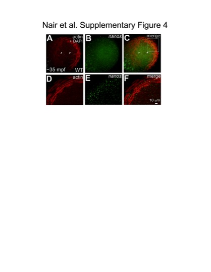
Fig. S4 Germ plasm RNPs localize onto cortical microfilaments at the blastodisc periphery. Animal views of blastodiscs; D–F are higher magnifications of area indicated in C, rotated 90° counterclockwise. Fluorescent in situ hybridization for the germ plasm mRNA nanos (B, E), together with immunolabeling for f-actin (A, D) shows that RNPs labeled with nanos co-localize with microfilaments arranged in concentric rings at the cortical periphery (C, F).
Acknowledgments
This image is the copyrighted work of the attributed author or publisher, and
ZFIN has permission only to display this image to its users.
Additional permissions should be obtained from the applicable author or publisher of the image.
Full text @ PLoS Genet.