
|
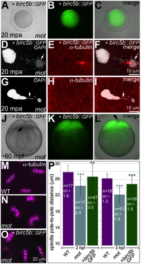
Fig. 3 Wild-type birc5b rescues cell division defects in motley mutants.
In vitro cultured oocytes from homozygous motley females express the injected birc5b::eGFP, mature and water-activate normally (A–C). Birc5b::eGFP-expressing motley eggs successfully complete meiosis as seen by the presence of meiotic midbody (E, F) and a distinctive polar body (D, F arrow). Control uninjected motley eggs do not have a meiotic midbody and form chromosomal bridges (G, I arrow) between condensed DNA masses (G–I). Normal cleavage furrows at ~60 mpf in in vitro fertilized embryos from Birc5b::eGFP-expressing eggs (J–L, arrows). Normal mitotic spindles in wild-type embryos (M), bent spindles in motley mutants (N), and mitotic spindles in Birc5b::eGFP-expressing motley embryos resembling wild-type (O). Due to the bending of the spindle, linear spindle pole-to-pole distance (SPD) is shorter in motley mutants than wild-type, while in Birc5b::eGFP-expressing motley embryos this distance approaches wild-type (P). Double-headed arrows in M-O indicate example SPD distances used for quantitation in P. Error bars in P represent Standard Deviation. Statistical significance of SPD values was ascertained by t-test (2-tailed, unpaired, unequal variance). P<0.001 = ***, P<0.01 = **. SPD distances were compared between uninjected motley and wild-type at 2 and 3 hpf (light blue asterisks), and between uninjected motley and birc5b::GFP injected motley at 2 and 3 hpf (green asterisks). P values were not significant (P>0.05), between wild-type and rescued birc5b::GFP injected SPDs.