
|
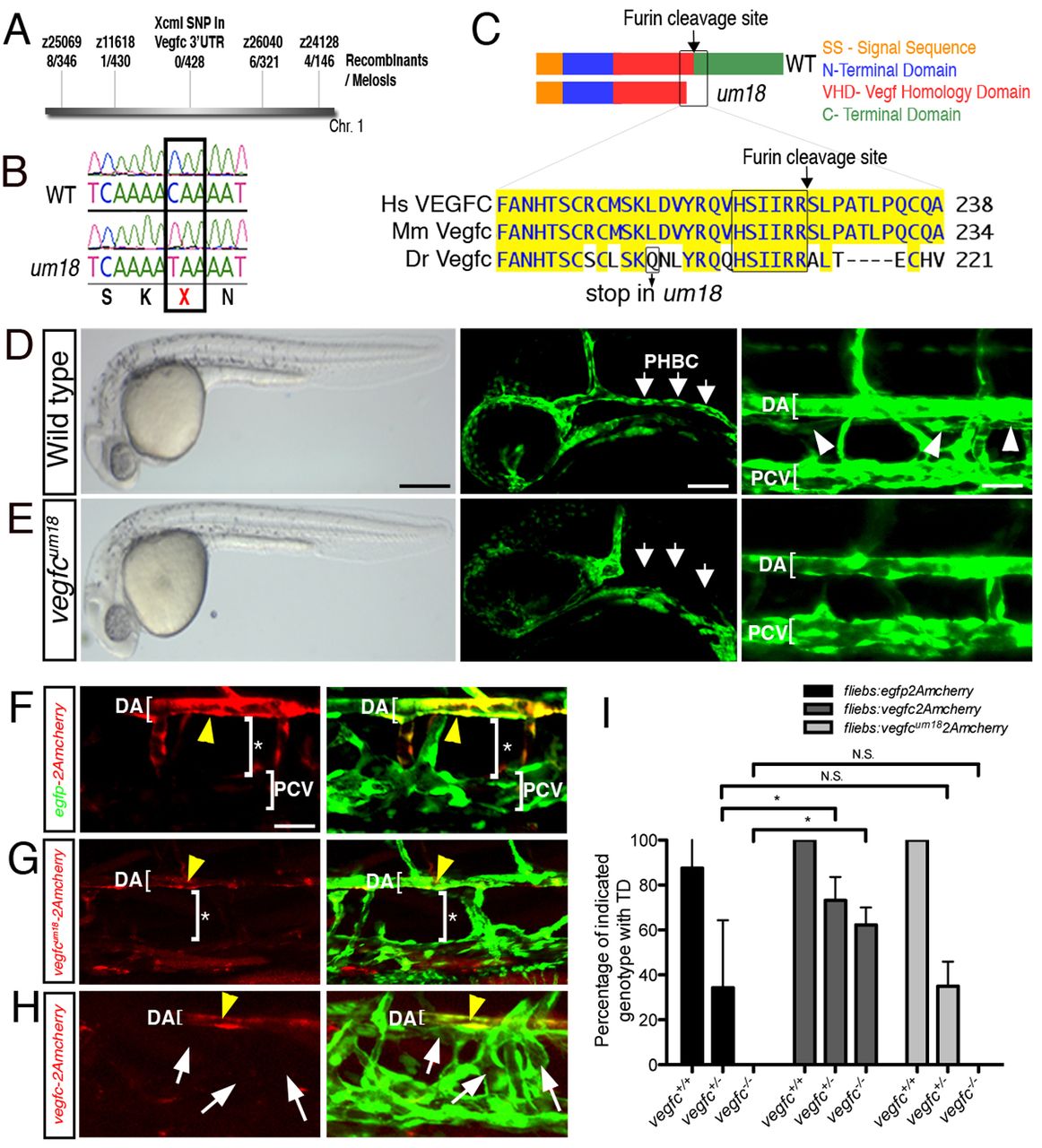
Fig. 1 vegfcum18 mutants display defects in vein and lymphatic vessel development. (A) Linkage map of markers on zebrafish chromosome 1 used in this study. (B) um18 mutation in vegfc exon 4. Box indicates the Gln202 to stop transition. (C) Vegfc domains and amino acid alignment near the um18 truncation. Hs, human; Mm, mouse; Dr, zebrafish. (D,E) (Left) Transmitted light images of embryos at 30 hpf. (Middle) Confocal micrographs of head blood vessels at 30 hpf. Primordial hindbrain channel (PHBC) is indicated by arrows. (Right) Confocal micrographs of trunk vessels at 5 dpf. Thoracic duct (TD) is indicated by arrowheads. Wild-type (D) and vegfcum18 mutant (E) Tg(fli1a:egfp)y1 siblings are shown. (F-H) Confocal micrographs of Tg(fli1a:egfp)y1 vegfcum18 mutant embryos co-injected with 25 pg tol2 transposase mRNA and 25 pg (F) pTol2-fli1ebs:egfp-2Amcherry, (G) pTol2-fli1ebs:vegfcum18-2Amcherry or (H) pTol2-fli1ebs:vegfc-2Amcherry. Yellow arrowheads indicate arterial endothelial cells expressing mCherry. Bracket with asterisk indicates absence of TD. White arrows indicate TD rescue. (D-H) Lateral view, anterior to the left, dorsal up. (I) Percentage of embryos showing TD of the indicated genotype injected with the indicated constructs. Values are the average of three independent experiments. *P<0.05; N.S., not significant; error bars indicate s.e.m. DA, dorsal aorta; PCV, posterior cardinal vein. Scale bars: 250 μm in D,E left; 50 μm in D,E middle; 25 μm in D,E right, F-H.