
|
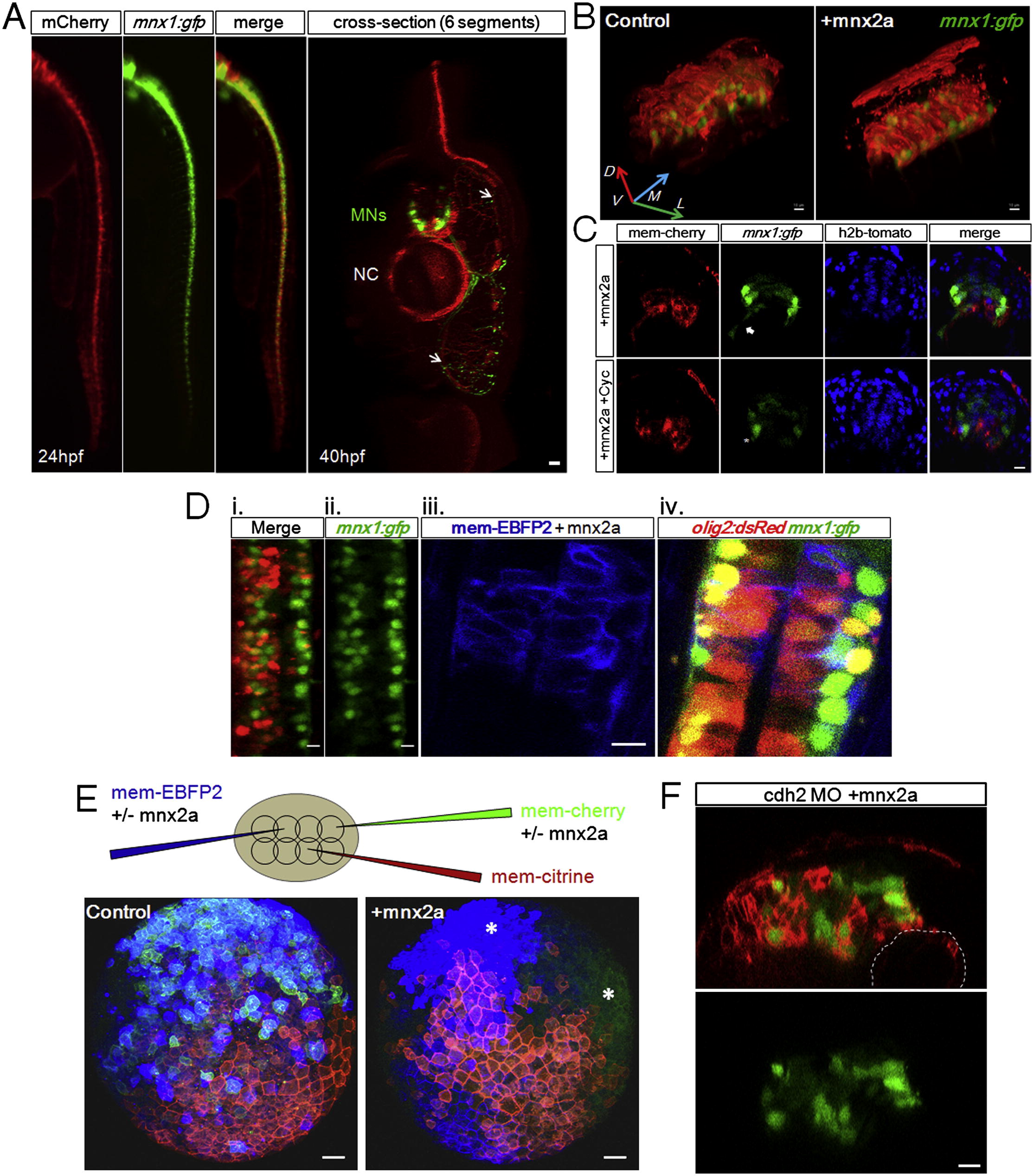
Fig. S6 Ectopic Mnx2a-Expressing Cells Populate pMN Domain in a Shh-Independent Manner, Mnx2a-Injected Cells Exhibit Changed Adhesion Properties, Related to Figure 6(A) Mnx2a injected cells localize to pMN domains and become MNs. Left: Lateral view of a ClassI embryo, the Mnx2a+mCherry coinjected cells predominantly segregated to the ventral neural tube, overlapping with the transgenic expression domain of mnx1:gfp. Right: mCherry+ cells become MNs. Arrows: mCherry+ motor axons in the muscle tissue. All scale bars (except E) are 10 μm.(B) Mnx2a cells populate extensively and exclusively the ventral neural tube. Images are 3D projections of original 2-channel z-stacks of 24hpf embryos. Unlike control embryos whose injected cells are randomly scattered in the neural tube (left), mnx2a injected cells form a clear ventral domain (right). The mCherry+ patch of cells seen above the neural tube are skin cells.(C) Cyclopamine treatment does not alter the Mnx2a phenotypes. 100 μM Cyclopamine was applied to dechorionated tg(mnx1:gfp/actb2:h2b-tdTomato) embryos at 7hpf. The axonal projections of the MNs were affected (Arrow, asterisk; Charron et al., 2003) but the pMN domain “stripe” formed by Cherry+ cells was not affected.(D) Mnx2a injected cells become pMNs. (i, ii). Top view of example tg(mnx1:gfp) embryo injected with Mnx2a and H2B-mCherry mRNA and treated with Cyclopamine at 12hpf, whose left half is populated by mCherry+ cells and have increased number of GFP+ cells compared to the right half. (iii, iv). Top view of example tg(mnx1:gfp/olig2:dsRed) embryo injected with Mnx2a and mem-EBFP2 mRNA. EBFP2+ cells are dsRed+ and show progenitor location and morphology. (E) Mnx2a injected cells form clusters at early neural plate stage. 3D projections (animal/dorsal view) of typical 8hpf triple injected embryos. Injections are performed as shown in the illustration. The formation of large clusters by Mnx2a cells as compared to random mixing of control cells suggests changes of adhesion properties in these cells, which may play a role in cell sorting. The injection amount of Mnx2a mRNA is higher (20ng/μl) in these experiments compared to Figure 6. Scale bar: 50 μm.(F) Mnx2a and cdh2 MO coinjected cells still form clusters and localize ventrally despite strong morphogenesis defects. Injections are performed as in Figure 6A. Many embryos show severe early defects and the survivors often show distorted neural tubes (image is a typical example). Mnx2a induced adhesion still appears to be in effect in these embryos. Dashed lines: Notochord boundary.
Reprinted from Cell, 153(3), Xiong, F., Tentner, A.R., Huang, P., Gelas, A., Mosaliganti, K.R., Souhait, L., Rannou, N., Swinburne, I.A., Obholzer, N.D., Cowgill, P.D., Schier, A.F., and Megason, S.G., Specified neural progenitors sort to form sharp domains after noisy shh signaling, 550-561, Copyright (2013) with permission from Elsevier. Full text @ Cell