
|
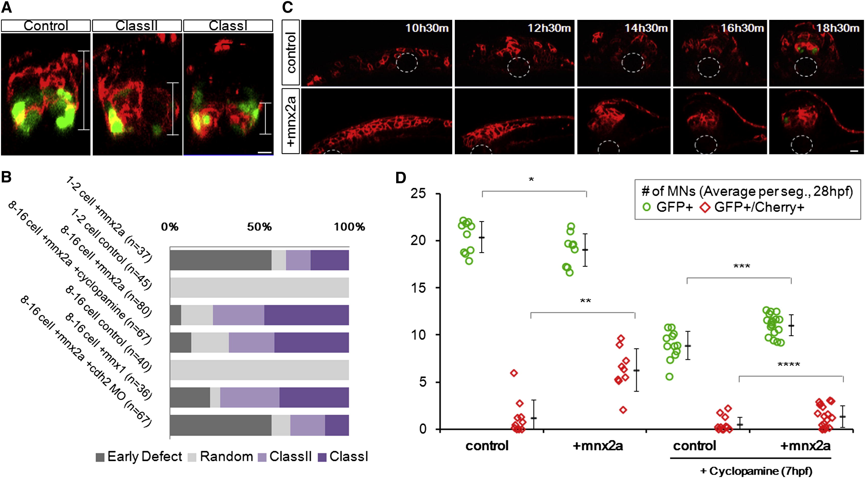
Fig. 6 Ectopic Mnx2a-Expressing Cells Form a Sharp Ventral Domain Similar to the pMN Domain (A) The 24 hpf neural tube phenotypes after injection of mem-mCherry α mnx2a mRNAs in one blastomere at 8- to 16-cell stage are shown. Phenotypes are classified according to the distribution of mCherry+ cells (brackets): class I embryos contain cells only in the ventral third of the neural tube, class II embryos contain cells in the ventral two-thirds, and “random” contains injected cells throughout. Green indicates mnx1:gfp. Scale bars, 10 μm. See also Figure S6A.(B) Summary of mosaic injection experiments. Early defect embryos failed to form neurula. Cyclopamine treatment started at 7 hpf. (C) Sample time course of Mnx2a domain formation. This Mnx2a embryo became class II type. Dashed-line circles indicate position of the notochord. Green shows mnx1:gfp. Red designates mem-mCherry. Scale bar, 10 μm. See also Figure S6B. (D) Mnx2a-expressing cells replace “normal” pMNs. Imaging and counting of MNs as Figure 3C. p = 0.09, p = 0.00004, p = 0.0001, and p = 0.03 (Student’s t test).
Reprinted from Cell, 153(3), Xiong, F., Tentner, A.R., Huang, P., Gelas, A., Mosaliganti, K.R., Souhait, L., Rannou, N., Swinburne, I.A., Obholzer, N.D., Cowgill, P.D., Schier, A.F., and Megason, S.G., Specified neural progenitors sort to form sharp domains after noisy shh signaling, 550-561, Copyright (2013) with permission from Elsevier. Full text @ Cell