
|
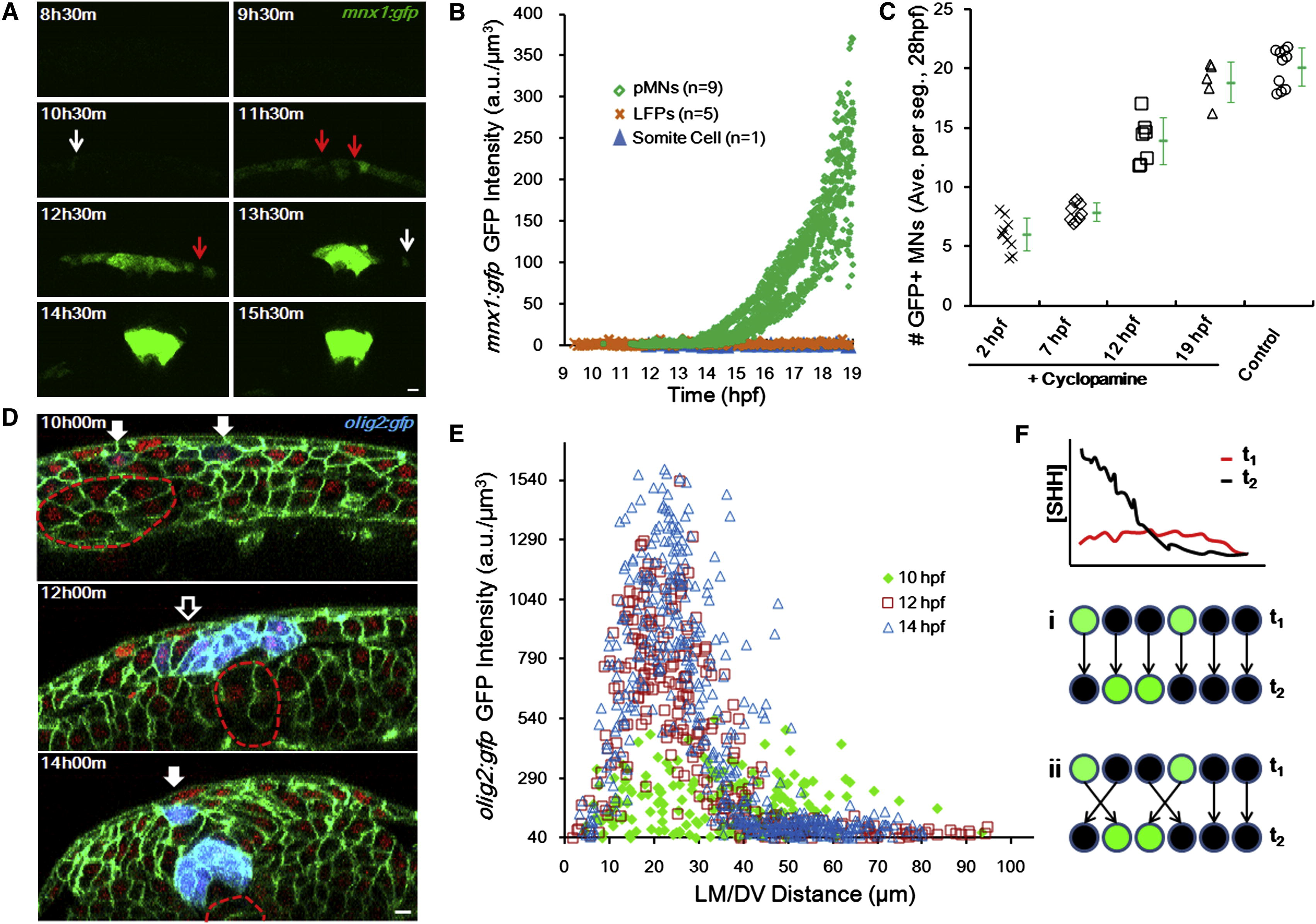
Fig. 3 Progenitor Fates Are Specified during Cell Movements in Mixed Distributions (A) Time course of mnx1:gfp expression. Images are cross-sectional examples. Red arrows point to mixed negative cells. White arrows indicate scattered positive cells. Scale bar, 10 μm.(B) GFP (mnx1:gfp) levels in tracked cells through time. See alsoFigure S3 A. (C) Time course of Cyclopamine inhibition of pMN specification. Treatment of 100 μM Cyclopamine started at indicated times, and MNs were counted at 28 hpf as an indicator of pMN number. Numbers are averaged per embryo by number of neural segments counted. Green marks show average (±SD). See also Figure S3 B. (D) olig2:gfp (blue) domain formation. Green indicates cell membrane. Red shows cell nucleus. Filled arrows point to scattered positive cells. Empty arrows indicate mixed negative cells. Dashed lines represent notochord boundary. Scale bar, 10 μm. (E) Spatial distribution of olig2:gfp+ cells. At 10 hpf, they scatter in a wider range and are mixed with negative cells; in contrast, at 14 hpf, positive cells form a major stripe between 15 and 30 μm where negative cells are absent. (F) Two models for sharp stripe formation. (i) Late (improved) gradient rewrites responses, predicting late specification and stable positions. (ii) Cell sorting corrects wrong positions, predicting early specification and rearrangement afterward.
Reprinted from Cell, 153(3), Xiong, F., Tentner, A.R., Huang, P., Gelas, A., Mosaliganti, K.R., Souhait, L., Rannou, N., Swinburne, I.A., Obholzer, N.D., Cowgill, P.D., Schier, A.F., and Megason, S.G., Specified neural progenitors sort to form sharp domains after noisy shh signaling, 550-561, Copyright (2013) with permission from Elsevier. Full text @ Cell