
|
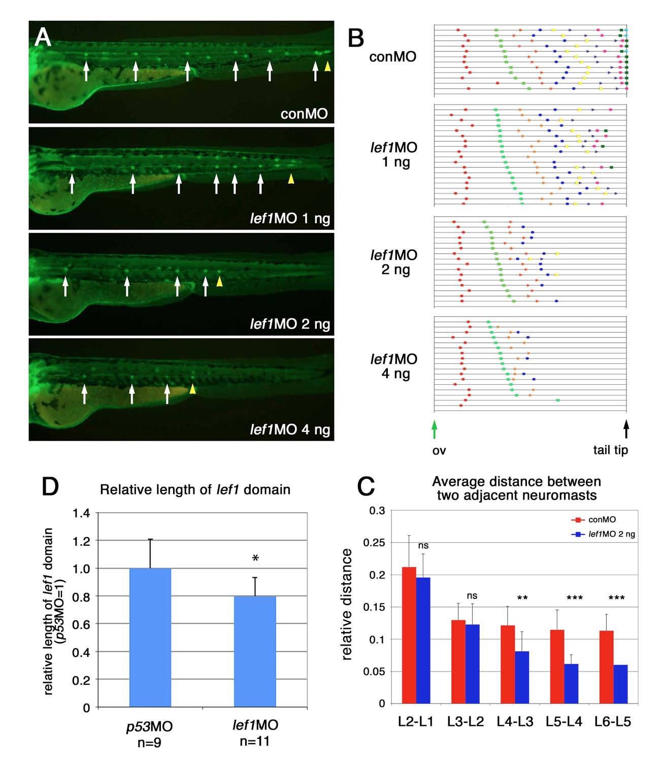
Fig. S5 Dose-dependent effects of lef1 knockdown on the pattern of neuromast deposition and PLLp termination. (A) Patterns of neuromast deposition at 48 hpf in embryos injected with 1, 2, 4 ng of lef1-MO. White arrows indicate deposited neuromasts and yellow arrowheads indicate the end of the PLLp migration. (B) Patterns of neuromast deposition in individual embryos injected with control-MO or 1, 2, 4 ng of lef1-MO. Individual embryos are represented on separate lines in an order determined by the relative position of L2. (C) Distance of two adjacent neuromasts deposited in embryos injected with control-MO (red) or 2 ng lef1-MO (blue). Data are presented as the average ± s.d. Statistical significance of the differences between each pair of groups was determined by Student’s t-test. **P<0.01, ***P<0.001; ns, not significant. (D) Effect of lef1-MO on the relative length of the leading lef1 domain at 31 hpf. Data are presented as the average ± s.d. Statistical significance of the differences between each pair of groups was determined by Student’s t-test. *P<0.05.