
|
Fig. S3
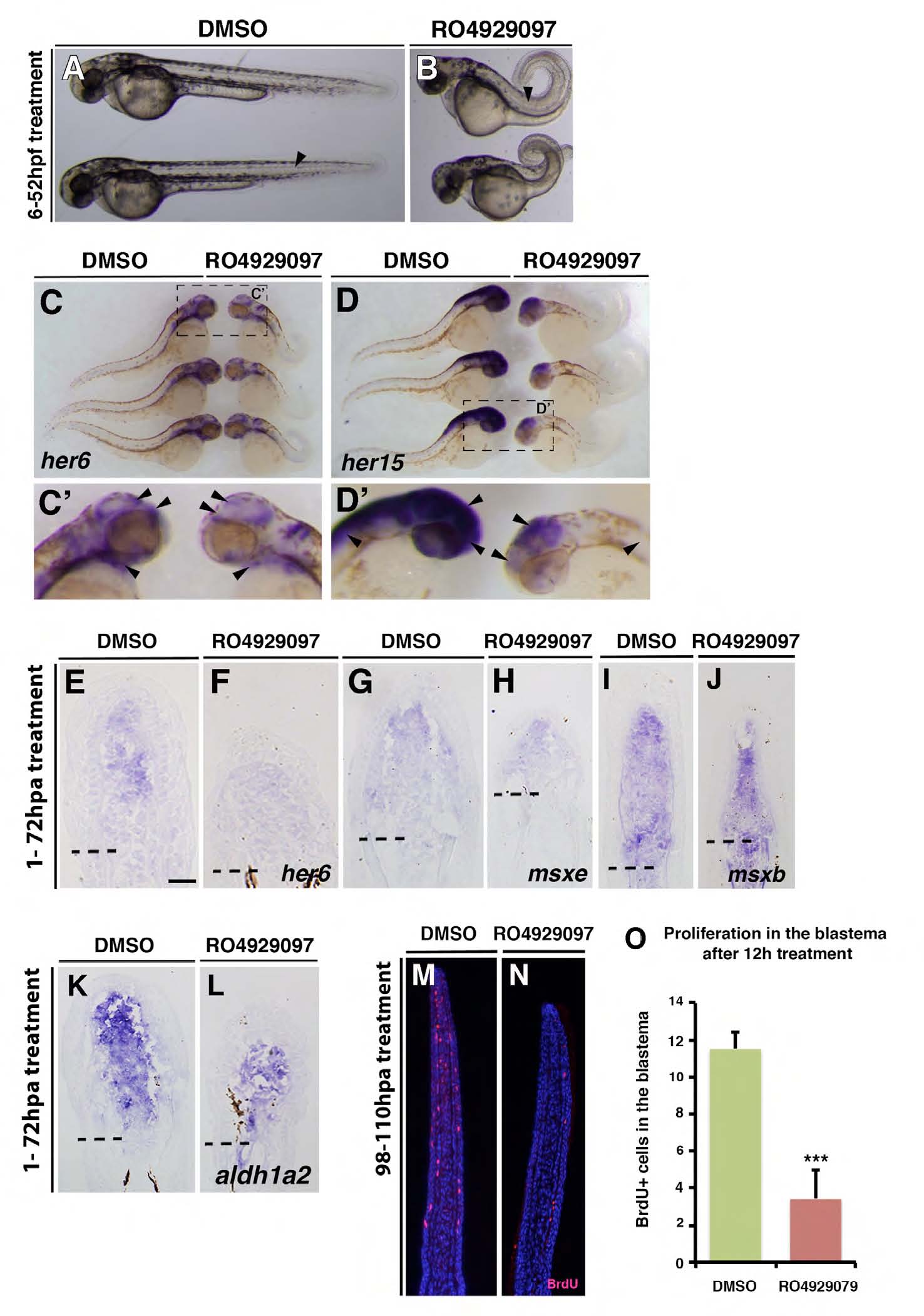
RO929097 treatment leads to Notch signalling knockdown in embryos and regenerating fins and reduces proliferation. (A-D) Embryos treated with either DMSO or 10 μM RO929097 from 6 to 52 hpf. RO929097-treated embryos show defects in somitogenesis (arrow) and a looped tail (B). (C,D) Whole-mount in situ hybridization: her6 gene expression is reduced in the brain and the gill mesenchyme of RO929097-treated embryos (arrowheads). her15 gene expression is reduced in the brain and spinal cord of RO929097-treated embryos (arrowheads). (E-L) In situ on fin sections after 72 hours of DMSO or 10 μM RO929097 treatment. her6 gene expression is reduced in RO929097-treated fins (E,F), but msxe (H), msxb (J) and aldh1a2 (L) expression seems to be unchanged in RO929097-treated compared with DMSO-treated fins (G,I,K). (M-O) Anti-BrdU-stained fin sections and quantification of BrdU+ cells within the distal most 300 μm of the mesenchyme. RO4929097-treated fins (n=4) (N) exhibit fewer BrdU-labelled cells than DMSO-treated fins (M) (n=4). ***P<0.001.