
Image
|
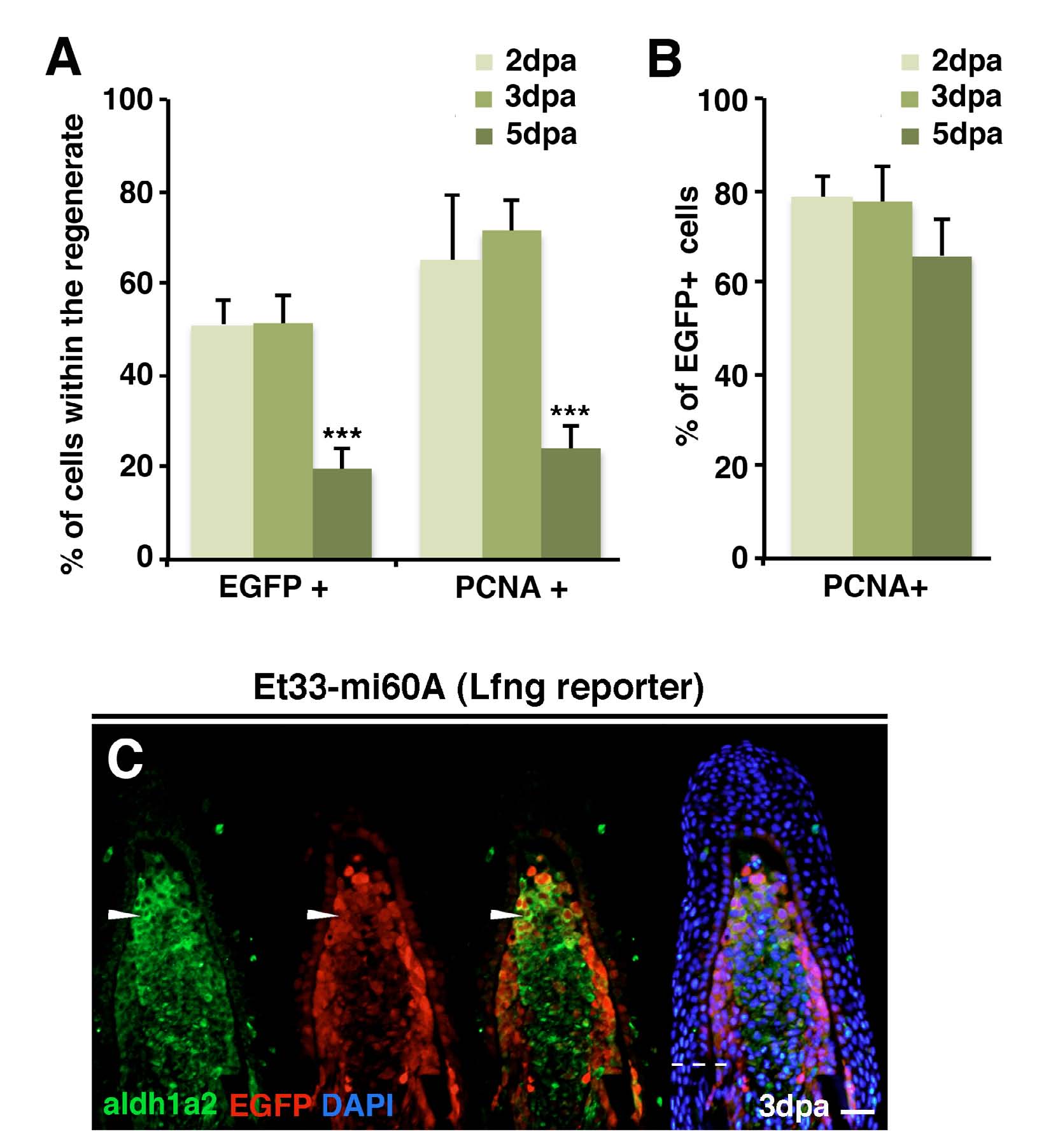
Figure Caption
Fig. S2
Lunatic-fringe-mediated Notch signalling is activated in proliferating, aldh1a2-expressing cells. (A) Mean percentage of EGFP-expressing cells in the regenerate of ET33-mi60A fin sections at 2 dpa, 3 dpa and 5 dpa. ***P<0.05. (B) Mean percentage of EGFP+ blastema cells co-labelled for PCNA in fin sections at 2 dpa, 3 dpa and 5 dpa. (C) Representative immunhistochemistry for EGFP and aldh1a2 in a 3 dpa fin section. Cells are double positive in the distal region of the blastema (arrowhead). Scale bars: 100 μm in C. Broken line marks the amputation plane.
Acknowledgments
This image is the copyrighted work of the attributed author or publisher, and
ZFIN has permission only to display this image to its users.
Additional permissions should be obtained from the applicable author or publisher of the image.
Full text @ Development