
|
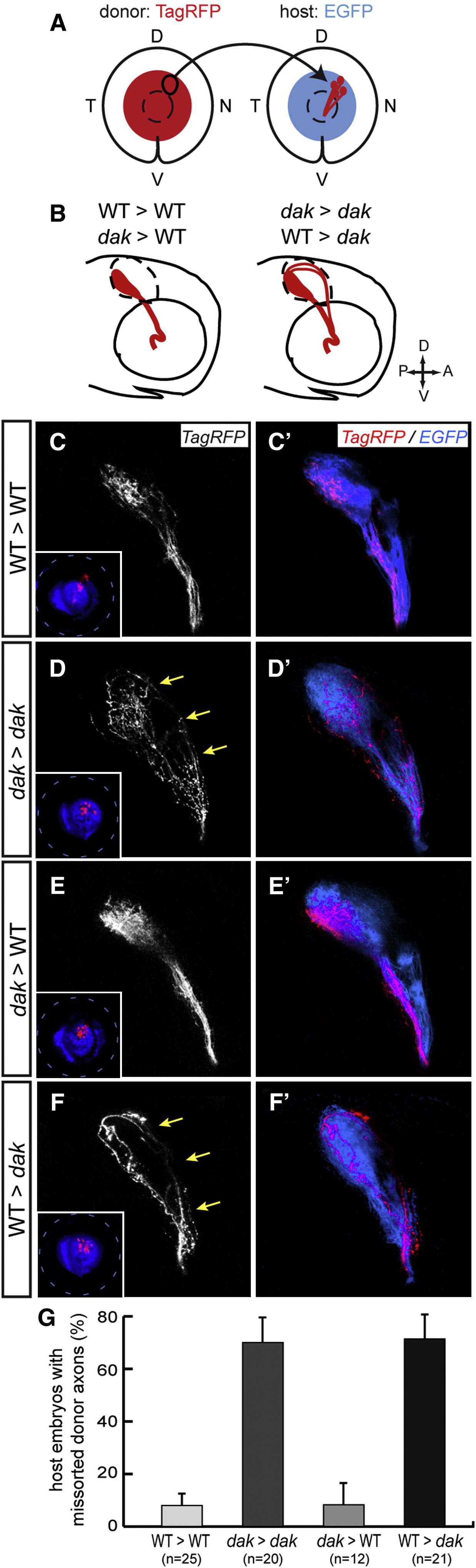
Fig. 4 HS Is Required Non-Cell-Autonomously for Correct Sorting of DN Axons (A) DN RGCs from an isl2b:TagRFP donor embryo were topographically transplanted between 30 and 34 hpf into the DN retina of an isl2b:EGFP host embryo. EGFP is shown in blue in the figure for the best visualization. (B–F′) Diagrams (B) and pictures (C–F′) of corresponding DN donor axons observed at 4 dpf. Lateral views, confocal maximal projections. In WT > WT (C and C′) and dak > WT (E and E′) transplants, DN donor axons (TagRFP+) pass through the ventral branch of the optic tract. In contrast, in dak > dak (D and D′) and WT > dak (F and F′) transplants, some donor axons are missorted along the dorsal branch of the tract (arrows) before reaching the tectum. Insets show transplanted DN TagRFP+ RGCs localized in the DN part of the host retina. (G) Quantification of missorting in transplants. The proportion of hosts with missorted donor DN axons was scored at 4 dpf. Missorting is observed in dak mutants but not WT hosts, indicating that HS is required non-cell-autonomously for correct sorting of retinal axons. Error bars correspond to SEs.