
Image
|
Figure Caption
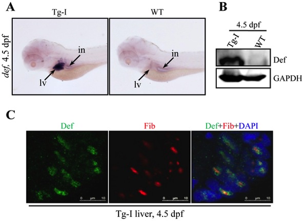
Fig. 2 Tg-I expresses high level of def specifically in the liver.
(A) WISH analysis of def expression in Tg-I and wildtype (WT) embryos at 4.5 dpf using the full length def cDNA as a probe. lv: liver, in: intestine. (B) Western blot of Def in Tg-I and wildtype (WT) embryos at 4.5 dpf. GAPDH: loading control. (C) Immunostaining of Def (green) and the nucleolar marker Fibrillarin (Fib) (red) in the liver of Tg-I fish at 4.5. DAPI was used to stain the nucleoli.
Figure Data
Acknowledgments
This image is the copyrighted work of the attributed author or publisher, and
ZFIN has permission only to display this image to its users.
Additional permissions should be obtained from the applicable author or publisher of the image.
Full text @ PLoS One