
|
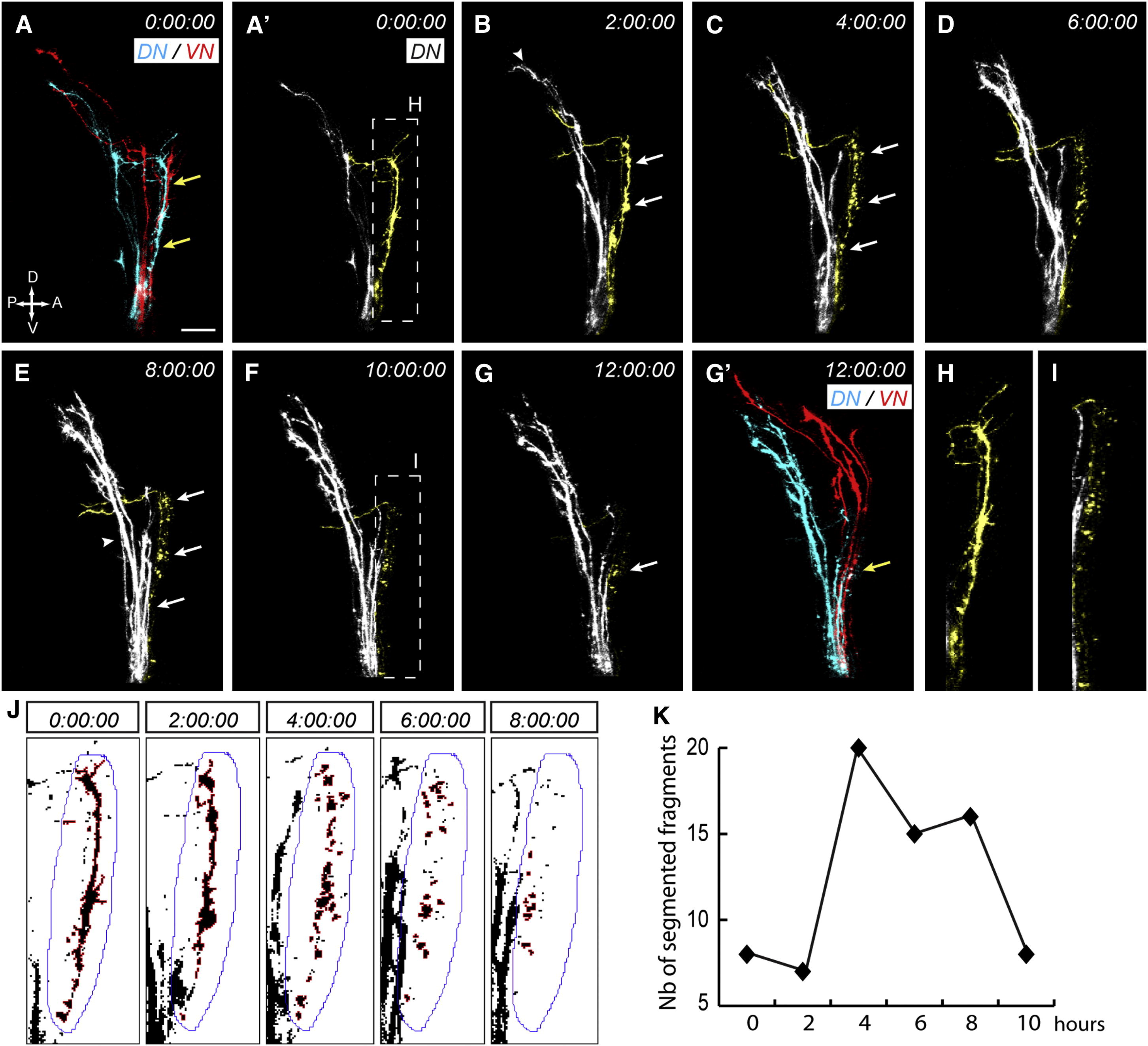
Fig. 2 Missorted Dorsal Axons Selectively Degenerate (A–G′) DN (blue, DiD) and VN (red, DiO) axons were topographically labeled at 48 hpf and time-lapse imaged at 15 min intervals for 12 hr starting at 54 hpf. The organization of DN and VN axons is shown at the beginning (A) and the end (G′) of the time-lapse, while time series of confocal image stack projections (A′–G) show DN axons only. Missorted DN axons are pseudocolored in yellow for the best visualization. Times indicate hours. Some DN axons are clearly missorted at 54 hpf (A′) and have a normal morphology (H). After 2 hr (B), correctly sorted axons have reached the tectum (arrowhead), while missorted DN axons stop their growth and start blebbing (arrows). They then fragment uniformly along their length over time (C–G, arrows), while correctly sorted axons (arrowhead in E) continue to grow toward the tectum. Fragments appear smaller at later stages (F, zoomed picture in I) and become hard to detect after 12 hr (G). Scale bar represents 20 μm. (See also Movies S1 and S2). (J) Maximal intensity projections of DiD signals at 0, 2, 4, 6, 8, and 10 hr (data not shown) were converted to binary images and a region of interest (ROI, blue line) surrounding missorted DN axons was defined. Axon segments within this region were then selected and analyzed using the “Analyze Particles” option in ImageJ. (K) Quantification of the number of axon fragments over time. Between 0 and 2 hr, the number of fragments is low, indicating that missorted axons are segmented as one main continuous region. The number of fragments then increases abruptly between 2 and 4 hr as axons degenerate and fragment. It then gradually decreases between 4 and 10 hr as axon fragments are progressively cleared. See also Figures S2 and S3.