
|
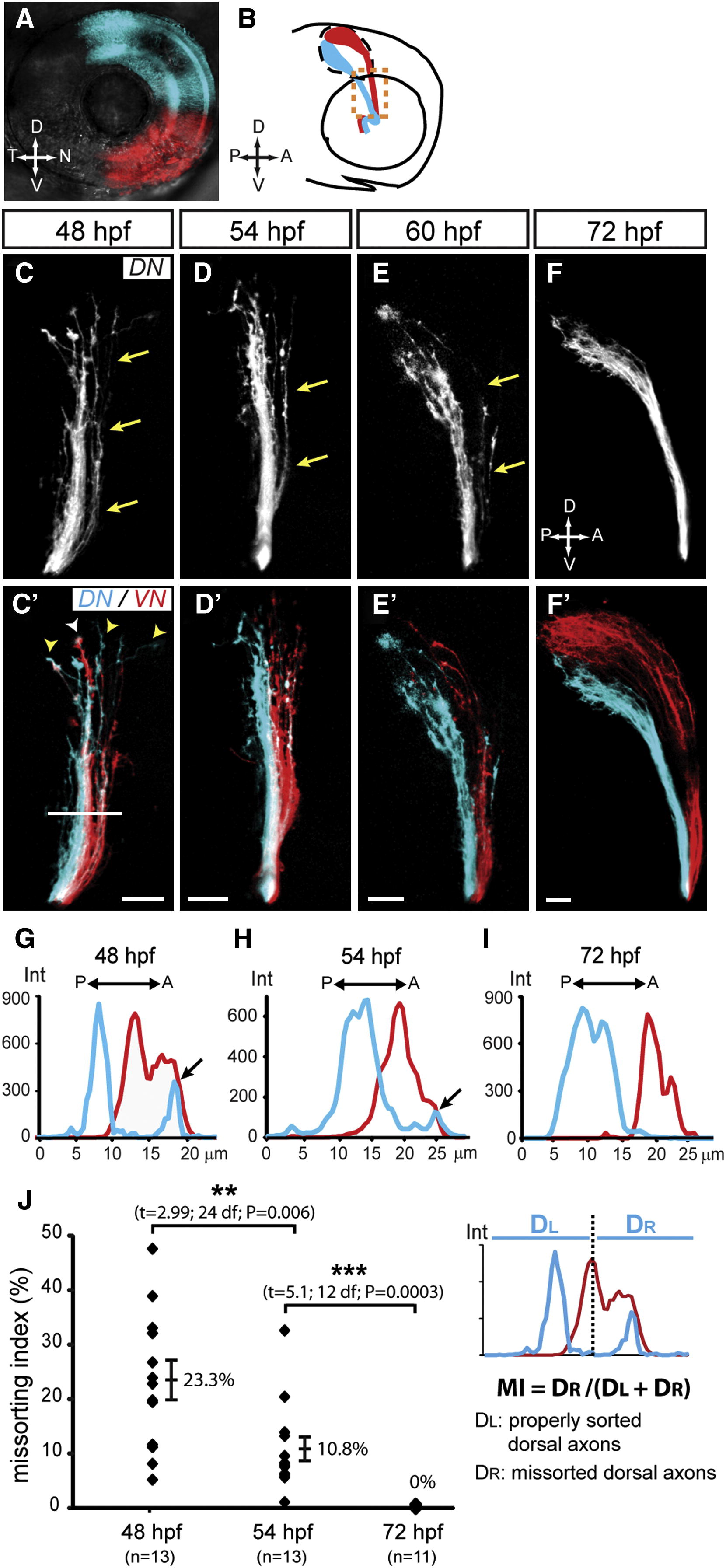
Fig. 1 Sorting of Retinal Axons in the Optic Tract Is Achieved through a Correction Mechanism (A) DiD (blue) and DiI (red) were topographically injected into the DN and VN quadrants of the retina, respectively. Lateral view, confocal maximal projection. (B) Diagram of corresponding DN (blue) and VN (red) retinal projections observed at 72 hpf in a lateral view after removing the contralateral eye. The optic tract region is demarcated with orange dashed line. (C–F′) Dorsal axon trajectories are progressively corrected between 48 and 72 hpf. At 48 hpf, DN (C, blue in C′) and VN (red in C2) axons are not precisely sorted in the optic tract. Some DN axons (arrows) are observed along VN axons in the most anterior/dorsal part of the tract. DN (yellow arrowheads in C′) and VN (white arrowhead) growth cones leading axons are intermingled. At 54 hpf (D and D′) and 60 hpf (E and E′), some DN axons are still missorted along VN axons (arrows). This missorting is no longer observed at 72 hpf (F and F′). Lateral views, confocal maximal projections; scale bar represents 20 μm. See also Figure S1. (G–I) Representative examples showing measurement of axon sorting in the tract at 48 hpf (G), 54 hpf (H), and 72 hpf (I). Intensity (Int, y axis) of DiD (blue) and DiI (red) signals was measured along a reference line drawn perpendicular to the tract (as indicated in C′). DiD signals are observed anteriorly to DiI signals at 48 and 54 hpf (arrows), but not at 72 hpf. (J) Quantification of missorting: a missorting index (MI) was calculated as the ratio of the signal intensity corresponding to missorted DN axons (DR) to the signal intensity of all DN axons (DL + DR). DN axons were considered missorted when located anterior to the VN axons with the highest signal intensity. Missorting of DN axons is evident at 48 hpf and becomes progressively corrected by 72 hpf. Error bars correspond to SEs. p < 0.05, p < 0.001.