
Image
|
Figure Caption
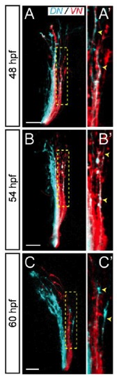
Fig. S1
Some ON axons are missorted at early stages of development(related to Figure I).
(A-C) ON (blue) and VN (red) retinal axons are not precisely sorted along the optic tract between 48 and 60 hpf: some ON axons are missorted along the dorsal branch of the tract (demarcated with yellow dashed line). (A′-C′) Zoomed views of the dorsal branch of the tract showing missorted ON axons (arrowheads). Lateral views, confocal maximal projections.
Acknowledgments
This image is the copyrighted work of the attributed author or publisher, and
ZFIN has permission only to display this image to its users.
Additional permissions should be obtained from the applicable author or publisher of the image.
Full text @ Neuron