
|
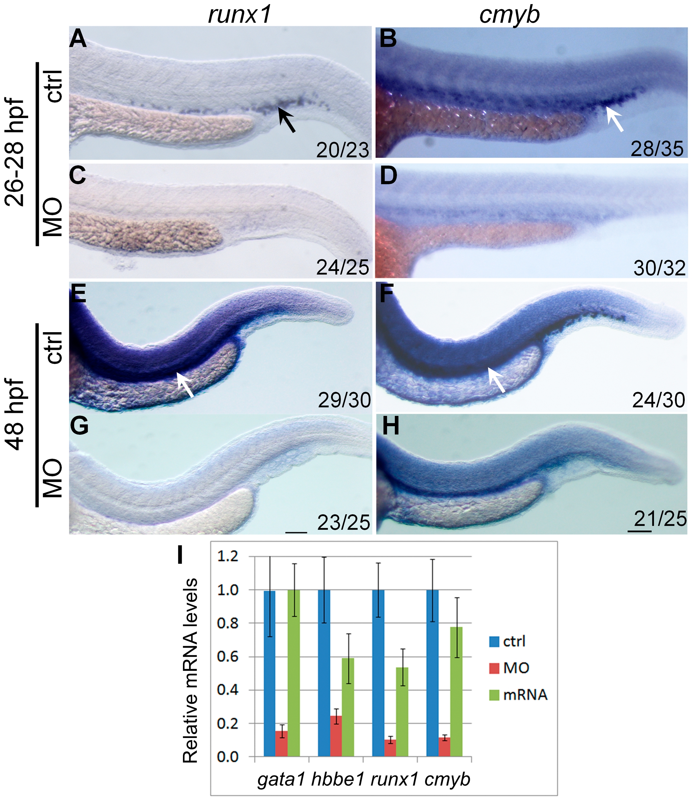
Fig. 3
hoxd4a expression is required for transient and definitive hematopoiesis.
In situ hybridization on 28 hpf (A–D) and 48 hpf (E–H) embryos showing expression of runx1 (A,C,E,G) and cmyb (B,D,F,H) in presumptive HSCs arising in the PBI (arrows in A and B) and AGM (arrows in E and F). Expression of both genes was severely reduced in hoxd4a morphants (C,D and G,H). Ratios in the bottom right corner of images indicate the fraction of embryos showing the presented phenotype. ctrl, embryos injected with a non-specific morpholino. MO, embryos injected with the anti-hoxd4a morpholino. hoxd4a mRNA, embryos simultaneously injected with the anti-hoxd4a MO plus capped mRNA for hoxd4a. Scale bars equal 100 μm. All images are at the same magnification. (I) qRT-PCR confirms the strong depletion of hematopoietic gene expression in hoxd4a morphants at 26–28 hpf, and restored expression following co-injection with capped mRNA for hoxd4a. Samples were normalized to β-actin. Error bars indicate standard error. By comparison to controls and rescuants, the gene expression levels of all morphants were statistically different to pd0.02 except for gata1 control vs morphant (p = 0.04) and hbbe1 rescuant vs morphant (p = 0.09).